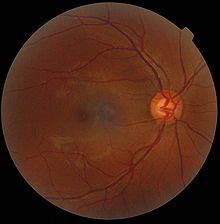
Blind spot (eye)


(pigment cell layer here dark red )
In ophthalmology, the blind spot is the point of the visual field onto which the exit point of the optic nerve in the outer space is projected.
Since there are no light receptors of the retina here, on the discus nervi optici with the optic nerve head papilla , this point represents a “blind spot” for the corresponding visual field region and so physiologically shows an absolute local field loss. The optic nerve papilla is anatomically located about 15 ° towards the nose ( nasal ) of the fovea centralis , so the blind spot is noticeable in the visual field about 15 ° towards the temple ( temporal ) as a characteristic scotoma - but usually only after a detailed examination of the monocular visual field.
General
Usually the blind spot is not noticed as a blind spot.
On the basis of sensations of the surrounding retinal regions and on the basis of those from the corresponding regions of the retina of the other eye, the optic nerve outlet of which is not depicted in the same area in the binocular field of vision, as well as on the basis of memory images, the perception of what is seen is supplemented into one image that the visual field loss does not appear subjectively.
But the blind spot does exist, there where the axons of the retinal ganglion cells converge to form the optic nerve and leave the eyeball - there are no photoreceptors of the retina. This is because these nerve fibers run on the inside of the retina, close to the vitreous humor , and need to be bundled with a gap to exit.
This "impractical" at first glance construction is explained by the development of the human eye from the optic vesicle , which is in the ontogeny as a protrusion of the brain forms and sinks for double-walled optic cup. A development that is similar in all vertebrates and with the result that the first system of the light-sensitive sensory cells arises from a layer of the inner leaf, which is enclosed by the outer leaf of the eye cup, which, when pigmented, forms the shading light filter of a camera obscura .
In the further development steps, additional neuron populations are formed, which are deposited in cell layers on the inside. This creates a structure that is also known as the inversion of the eye : facing the outer pigment epithelium , the photoreceptors are now facing away from the light under the later inner layers of the retina , in particular the nerve fiber layer .
In some other living things, the eyes are structured differently. If, for example, the light-sensitive sensory cells originate from outer tissue layers of the surface ectoderm , then the eyes do not have such a blind spot. In cuttlefish, for example, the light-sensitive cells form the innermost layer of the retina facing the light. Their signals are passed on by afferent neurons that are located further outside and thus approach from the side facing away from the light. Their nerve cell extensions, which run as fibers of an optic nerve from the eye to regions of the brain, therefore do not have to pass through a gap in the photoreceptor layer of the retina.
Self-experiment
Instructions: cover the right eye and fix the X with the left eye . If the screen is about three times the distance between the two letters, the O can no longer be seen. Three times the distance is only a guideline. If the O can still be seen, vary the distance to the screen until the O disappears. Conversely for the blind spot of the right eye: cover the left eye, fix the O and the X disappears.
| Try to try it out | ||
|---|---|---|
| O | X | |
More self-illustration
Instructions: cover the left eye and fix the point with the right eye. Start with a screen distance of about twice the distance between the black point and the center of the white circle. If you then slowly increase the distance, you can see how the missing part of the pattern is completed, even if no “information” is transmitted via a visual cell. The incomplete information is completed by the brain. This process is called filling-in.
discovery
The blind spot was discovered in 1660 by the French naturalist Edme Mariotte . He presented his discovery at the French royal court by making a small coin seemingly magically "disappear" with an attempt - similar to the tests mentioned above.
See also
Individual evidence
- ↑ Hans-Werner Hunziker: In the eye of the reader: foveal and peripheral perception - from spelling to reading pleasure . Transmedia Stäubli Verlag, Zurich 2006, ISBN 978-3-7266-0068-6 .
- ↑ Stefan Pollmann: General Psychology . UTB, 2008, ISBN 978-3-497-01971-7 .
literature
- Axenfeld / Pau: textbook and atlas of ophthalmology. With the collaboration of R. Sachsenweger et al., Stuttgart: Gustav Fischer Verlag, 1980, ISBN 3-437-00255-4 .
