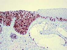
Carcinoma in situ
| Classification according to ICD-10 | |
|---|---|
| D00-D09 | In-situ neoplasms |
| ICD-10 online (WHO version 2019) | |
As carcinoma in situ (CIS) (literally "cancer at the source"), an early stage of an epithelial tumor called without invasive tumor growth, which only grows intraepithelial such. B. in the uppermost layer of skin or mucous membrane or in the milk ducts of the mammary gland . The individual cells cannot be distinguished microscopically ( histologically , immunohistologically ) in their cellular structures and their relationship to one another from those of an invasively growing carcinoma , but the basal lamina has not yet broken through, and there is no tumor invasion. The carcinoma in situ does not metastasize ; that is, it cannot form deposits in lymph nodes or other organs.
The importance of the CIS lies in the fact that it can develop into a locally invasive (malignant) tumor, but because of the different lengths of latency in individual cases, it can not be predicted when a CIS will break through the basement membrane. Even after incomplete excision, CIS can recur as an invasive carcinoma and then metastasize.
Examples

- Actinic cheilosis : CIS of the red lips
- Actinic keratosis : CIS of the skin, chronic damage to the cornified epidermis caused by long-term intensive exposure to sunlight ( UV radiation )
- Anal intraepithelial neoplasia stage III (AIN III) of a CIS of the anus
- Bowenoid papulosis mostly in men ( penile intraepithelial neoplasia stage III - PIN III): CIS of the penis
- Ductal carcinoma in situ : (DCIS), most common CIS of the breast
- Erythroplasia : CIS on the genitals ( penis , penis foreskin , labia ), skin disease caused by human papillomavirus (HPV) that can lead to skin cancer ( precancerosis )
- Lobular carcinoma in situ : (LCIS), second most common CIS of the breast
- Bowen's disease : CIS of the skin, HPV infection in the genital area, which leads to characteristic nodular (papular) skin changes
- Verrucous leukoplakia : CIS of the oral (mouth) mucous membrane or other internal mucous membranes ( larynx , esophagus , conjunctiva )
- Vaginal intraepithelial neoplasia stage III (VAIN III): CIS of the vagina
- Vulvar intraepithelial neoplasia stage III (VIN III): CIS of the vulva
- Cervical intraepithelial neoplasia stage III (CIN III): CIS of the cervix , more precisely the portio vaginalis
therapy
- CIS of the cervix
- During the gynecological check-up, the early detection of a CIS on the cervix (Portio vaginalis uteri) can prevent further development into cervical carcinoma , a real cancer. To do this, a cone from the cervix is operated on ( conization ), so the uterus is preserved.
- There are similar healing small interventions in other body tissues, for example colon polyps or in the breast.
- CIS of the skin or mucous membranes
- Ideally, CIS of the skin should always be removed with a sufficient safety margin. If this is not possible for cosmetic or functional reasons (extensive CIS on the face; genitals), immunomodulating ointment treatment with imiquimod , cryotherapy (freezing), local chemotherapy with 5-fluorouracil or radiation therapy are possible .
Histology and Chromosomal Changes
The histological picture correlates with chromosomal and genetic changes. In oral squamous cell carcinoma , for example, a normal epithelium first develops into a hyperplastic epithelium due to the inactivation of the p16 gene; the further development to the dysplastic epithelium occurs through the mutation of the tumor suppressor gene p53; the carcinoma in situ is characterized by the amplification of the cyclin D1 gene; In the case of invasive carcinoma, the inactivation of the PTEN gene can also be demonstrated. The loss of such tumor suppressor genes has been described for a large number of tumors.
Individual evidence
- ↑ Peter Fritsch: Dermatology and Venereology. 2nd Edition. Springer Verlag, 2004, ISBN 3-540-00332-0 .