
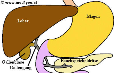
Common bile duct
The ductus choledochus (from Latin ductus "duct", choledochus "bile receiving"), also called the main bile duct , is the union of the ductus hepaticus communis with the duct of the gallbladder ( ductus cysticus ) and serves to transport the bile into the duodenum (duodenum) . In mammals that lack a gallbladder (e.g. horses ), the union of the right and left hepatic ducts is known as the common bile duct.
anatomy
The common bile duct is a membranous, muscular tube approximately 2-3 mm in diameter and 10 cm in length in humans. Together with the portal vein and the arteria hepatica propria, it passes through the porta hepatica . Then it runs over the small mesh behind the C-loop of the duodenum along the head of the pancreas . There it unites in humans with the main duct of the pancreas ( Ductus pancreaticus or Ductus wirsungianus after the German anatomist JG Wirsung ) and, after the passage of a sphincter muscle ( papilla vateri or papilla duodeni major ), opens on the inside of the C-loop of the duodenum. The common bile duct is supplied with blood via the cystic artery .
Function and meaning
When sober, the bile is backed up in the gallbladder. There the bile is collected and thickened a little. The gallbladder serves as a reservoir.
After eating, the flow reverses in the gallbladder duct. The gallbladder contracts, emptying the bile into the duodenum.
The flow velocity in the corridor is too slow to be recorded with an ultrasonic Doppler device. It is estimated at 0.1–1 centimeters per second. The different directions of flow are controlled by the sphincter muscles and by hormones, especially cholecystokinin .
Diseases
Permanent occlusion of the bile duct is a serious health disorder. At first there is jaundice and finally liver failure . A blockage of the bile duct must therefore be removed again through medical measures.
The occlusion of the common bile duct can be caused by a gallstone ( choledocholithiasis ), a bile duct tumor or external compression (e.g. pancreatic head tumor ). After a gallbladder operation, accidental injury or clamping (surgical clip) can lead to a complete closure.
Inflammation of the common bile duct can occur in the form of bacterial cholangitis (secondary sclerosing cholangitis) or as an autoimmune disease ( primary sclerosing cholangitis ). The resulting narrowing of the bile duct is mostly found in the area of the intrahepatic bile duct
A atresia can be used as congenital malformation occur. Malformations are also possible in the area of the mouth: Choledochus cyste (cystic expansion of the ductus choledochus), choledochocele (3 to 6 cm large cystic expansion of the ampulla vateri), inner duodenal diverticulum (protrusion of a residual membrane in the duodenum at the level of the papilla vateri).
Investigation options
The common bile duct can now be assessed quite well through a number of examination options. Most important are the ultrasound or endosonography , various laboratory values ( gamma-glutamyltransferase , bilirubin , alkaline phosphatase ) and endoscopic retrograde cholangiopancreatography (ERCP). Also, a percutaneous transhepatic cholangiography (PTC), computed tomography (CT) or magnetic resonance cholangiopancreatography (MRCP) are possible.
The previously common indirect representation of the duct using bile-permeable contrast media is hardly used today, since the ultrasound and the ERCP provide faster and better information and the previously more frequent contrast media intolerances are thus eliminated.
Ultrasound examination
The common bile duct can be visualized by both an abdominal ultrasound (through the skin) and an endosonography (from the inside as part of an endoscopic examination). The corridor is typically less than 0.7 cm. In the pathological state, it can swell up to 2 cm in width. After gallbladder surgery, a width of up to 1.1 cm is still normal, as the bile duct can partially take over the storage function of the gallbladder.
The common bile duct runs through the portal of the liver together with the hepatic artery (hepatic artery) and the portal vein . It runs parallel to the portal vein and is crossed several times by the hepatic artery. In order to distinguish the common bile duct from these two blood vessels, color-coded Doppler sonography (duplex sonography), which can show the blood flow, is helpful.
The duodenum is usually filled with air and can make it difficult to visualize the common bile duct (especially in the last section in the area of the pancreas ) from the outside. From the inside, however, the common bile duct can usually be visualized without any problems.
ERCP
A direct visualization of the common bile duct is possible with X-ray contrast medium by probing the papilla through an endoscope with side-view optics ( endoscopic retrograde cholangio-pancreatography , ERCP ). If there are abnormal findings, the therapy can be carried out in the same session. Stones can be removed from the duct or stenoses can be bridged with plastic or wire stents . Malformations such as choledochoceles and internal duodenal diverticula are also corrected endoscopically and surgically. The first endoscopic choledochocele cleavage was carried out by SE Miederer at the University of Bonn in 1976.
CT and MRI
If the ultrasound examination does not produce a conclusive result and there is still a clinical suspicion of a biliary tract disease - e.g. B. stone disease, tumor or malformation - an objectifiable, low-overlapping and multi-dimensional imaging is expedient. Computed tomography (volume scan with MPR), which is quickly available and inexpensive, but also means exposure to radiation, and MRI (especially MRCP), which is relatively expensive and can only be planned, are suitable for this. Both methods are only used to present findings. An option for intervention or therapy - as with ERCP - is only given here in exceptional cases.
literature
- ^ G. Schmidt: Ultrasound Companion . Thieme
- ↑ Miederer, SE et al .: Endoscopic transpapillary splitting of a choledochocele. Dtsch Med Wochenschr. 1978 Feb. 3: 103 (5): 216,219. PMID 631041
- ↑ Miederer, SE et al .: Choledochocele and Intraduodenal Diverticulum. Switzerland Rundsch Med Prax. 1981 Nov 10; 70 (46): 2068-76. PMID 6796952