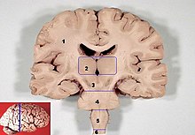
Medulla oblongata

The rhombencephalon of the 3-vesicle stage (left) differentiates into the metencephalon and the myelencephalon , from which the medulla oblongata arises, bordering the spinal cord.
The medulla oblongata , the elongated medulla or medullary brain , is the most caudal (back or bottom) part of the brain stem and belongs to the brain and thus to the central nervous system .

This area of the brain is anatomically also Myelencephalon called (Mark brain), other names are hindbrain and bulb medulla spinalis , bulb cerebral or Bulbärhirn . Together with the metencephalon (hindbrain), the myelencephalon forms the rhombencephalon ( hindbrain ). The medullary brain or elongated marrow is not sharply demarcated caudally in the transition to the spinal cord ( medulla spinalis ). By definition, it extends from the origin of the first pair of spinal nerves - approximately at the level of the foramen magnum - up to the bridge ( pons ) of the hindbrain; in adult humans it is about three inches long.
In the elongated medulla lie the vital centers for the regulation of breathing ( breathing center ) and the blood circulation and others, via which reflex reactions can be triggered: nutritive such as sucking reflex and swallowing reflex , but also protective such as coughing reflex , sneezing reflex , gag reflex and vomiting (see also Vomiting center ). Chemosensors can also be found in the medulla oblongata itself, for example for the acid-base status in the body. In addition, all the pathways that connect other areas of the brain, such as the cerebrum , to the spinal cord, descend through the medullary brain. Conversely, pathways ascending from the spinal cord, like those from the posterior cord , are switched in the medullary brain.
A failure of the medulla oblongata, for example in severe injuries to the cervical spine , usually leads to death. On the other hand, a person in whom only the cerebrum is largely or completely inoperable ( partial brain death ) can continue to physically live with the help of the functions regulated in the intact medulla oblongata. Since the centers for breathing are located here, such a patient does not need artificial ventilation - except in crises. The patients are in a deep coma and mostly show an apallic syndrome . If the upper brain stem is disturbed, it is referred to as a midbrain syndrome ; if the brain stem functions in the area of the medulla oblongata, it is referred to as a bulbous brain syndrome .
structure
In the case of the medulla oblongata or the myelencephalon, a slender lower section around the central canal, similar to the spinal cord, as well as - with its widening to the fourth cerebral ventricle - a broad upper section, which lies unfolded on the floor of the lower diamond-shaped pit , and a different structure can be distinguished shows. Here the gray matter takes up the entire width as a tegmentum (hood), is loosened up like a reticular formation like a network and contains numerous core areas embedded in it, including the cores of the cranial nerves (V, VII-XII) in a functional sequence from the center outwards.
Ventrally superimposed on the medullary hood is an area of white matter , in which mainly descending tracts run, and which externally forms a bulge on both sides of the anterior longitudinal furrow, called a pyramis . These downwardly slimmer pyramids are connected in the caudal area as decussatio pyramidum ( pyramidal crossing), where many nerve fibers cross from one side to the other, so most of the pyramidal tracts from the cerebral cortex for muscle movements. The median line between the two halves is therefore also known as the raphe (seam).
The motor XII is the lowest cranial nerve . Nervus hypoglossus in front from the medulla oblongata, between the pyramid and a lateral thickening, called oliva (olive). This contains the nuclei olivares (olive nuclei ) of the medullary brain, a core complex in which the main nucleus ( nucleus olivaris principalis ; also called "olive") and secondary nuclei ( nuclei olivares accessorii medialis et posterior ; also called "minor olives") are distinguished.
In the lower olive ( nucleus olivaris inferior ) in the medullary brain, projections from other brain areas and proprioceptive ones from the spinal cord are brought together and switched to the next neurons. From here, paths lead to the cerebellum , which plays a central role in the coordination of muscle movements, especially for fine motor movements.
literature
- Martin Trepel: Neuroanatomy . 5th edition. Urban & Fischer, Munich 2012, ISBN 978-3-437-41299-8 , pp. 115-136 .
- Franz-Viktor Salomon: nervous system, systema nervosum . In: Salomon / Geyer / Gille (Hrsg.): Anatomy for veterinary medicine . Enke, Stuttgart 2004, pp. 464-577. ISBN 3-8304-1007-7

