
Pirogoff amputation

The Pirogoff amputation (also amputation according to Pirogoff , amputation according to Spitzy , amputation according to Pirogoff-Spitzy or in English after Boyd) is a surgical method for amputating the foot , which was invented in 1854 by Nikolai Ivanovich Pirogow . It receives the heel with its cushion and is therefore capable of endurance. However, by removing the ankle bone and then stiffening it, the mobility of the rear foot is completely lost. A lower leg prosthesis is necessary for rehabilitation in order to obtain an appropriate abutment for the foot prosthesis.
purpose
If the soft tissues and bones of the foot are lost in a gangrene or injury, and the equinus foot position is more than 45 °, but the heel is still intact, the Pirogoff amputation is a good option. Without an equinus foot position, a disarticulation can be performed in the Chopart joint , which preserves the ankle joint. The next higher amputation, if the skin defect is too large, is the supramalleolar amputation according to Syme , in which the skin of the heel is also preserved and thus a stump that can withstand endurance, is about 4–7 cm shorter.
technology
The surgical access for the amputation is from the front (ventral), with the formation of a sole flap that extends further distally , which can then cover the ventral defect and also create an end load capacity there. The skin on the back of the foot, on the other hand, is very sensitive to pressure and endangered where pressure is created by the load or the prosthesis. After exposing the ankle bone, it is completely removed ( astragalectomy ). The articular surface of the upper ankle joint , together with the inner and outer ankle , is resected through a horizontal osteotomy with an oscillating saw, with particular care being taken to protect the vessels behind the inner ankle. This is followed by a horizontal osteotomy on the calcaneus, which removes all of the joint parts of the subtalar joint. By removing the ankle bone, about 3 to 4 cm of space is gained, so that even a sharp shortening of the Achilles tendon is usually compensated for and a tendon lengthening is rarely necessary. The malleoli are removed for a good prosthesis. The calcaneotibial arthrodesis takes place under compression and with an advance of the heel bone by about 10 to 15 mm, whereby the heel bone must be adjusted with a slight external rotation as on the opposite side. The distal-plantar edge of the calcaneus must then be rounded off, especially on the outside, in order not to create any pressure-sensitive bony prominence. As osteosynthesis a is mostly external fixator with two Steinmann nails or long screws in the tibia and calcaneus used alternatively crossed K-wires , which are inserted from the proximal end of the tibia and calcaneus thereby not hurt the sole of skin. A skin closure must take place without tension.
The permanent fixation by means of wires, screws or external fixators enables immediate loading of the residual limb after swelling and wound healing, and a prosthesis is provided after the postoperative edema has completely receded and the material has been removed after about six weeks.
Prosthesis supply
A sole shoe supply with a forefoot prosthesis is not sufficient, as it cannot be fixed firmly to the stump and therefore has no functional significance. There is also a risk of pressure points and abrasions. For this reason, a lower leg prosthesis is usually manufactured which, thanks to the lower leg-length frame, can transfer the weight to the prosthetic foot, especially at the edge of the shin, and thus enables a largely physiological gait pattern with the foot rolling. The lower leg frame can be manufactured as a front frame prosthesis, since it does not have to take on any weight in the case of a residual limb that is capable of bearing endurance, and the risk of pressure points is thus significantly reduced.
However, only the 4–5 cm shortening remains for the prosthetic ankle, which limits the scope for choosing the ankle. A joint is often dispensed with and a rigid prosthesis is made. Then, however, a roll-off cradle and a wedge heel on the shoe are necessary to enable the roll-off process.
Since the residual limb can withstand the ultimate load, mobility is also possible without a prosthesis, but with shortening limbs and without rolling, i.e. H. the gait costs significantly more power and is significantly less physiological.
Complications
In general, amputation stumps, especially with arterial occlusive disease, have a high risk of recurrence of circulatory disorders and necrosis and subsequent amputations. Rearfoot amputations are considered to be significantly more risky because the vascular supply is borderline and muscular coverage of the stump is difficult. Post-operatively, there is always a swelling that further compresses the blood supply.
There are recommendations not to perform a hindfoot amputation in the case of severe arterial occlusive disease, but rather to perform a proximal lower leg amputation, which usually heals more quickly, enables adequate muscular residual limb coverage and has a better blood flow situation.
The build-up of the arthrodesis, however, is not a problem and pseudarthroses are very rare.
photos
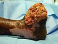
Leper foot

X-ray of the foot

X-ray AP after tibio-calcaneal arthrodesis (Pirogoff)

lateral admission
.JPG)


literature
- M. Schofer, M. Settner, HR. Kortmann: Amputations on the foot . In: Trauma and Occupational Disease. 3 (2001), pp. 244-247.
Web links
- Anton Hanauer: The amputation according to Pirogoff. In: Archive for orthopedic and trauma surgery, with special consideration of fracture theory and orthopedic-surgical technology . December 1923.
Individual evidence
- ↑ a b c d René Baumgartner , Pierre Botta: Amputation and prosthetic care of the lower extremity . Enke-Verlag, Stuttgart 1995, ISBN 3-432-97502-3 .