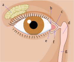
Lacrimal gland
The lacrimal gland ( Latin: Glandula lacrimalis ) is a gland that produces most of the tear fluid . The lacrimal gland lies laterally on top of the eye within the eye socket . It produces electrolytes and a large number of protein compounds. Its secretion is directed through 6 to 12 ducts into the vault of the conjunctival sac ( fornix conjunctivae ) and distributed over the cornea by blinking . The tear fluid keeps the cornea moist and nourishes it.
The lacrimal gland, together with the additional lacrimal glands producing the portion of the lacrimal system . The tear fluid that is formed is passed into the nasal cavity via the draining tear ducts .
histology
The human lacrimal gland is a purely serous gland with tubulo- acinous end pieces and, as such, can be differentiated from the parotid and pancreas in the light microscope specimen : a switching and strip system, in contrast to the parotid gland, is not formed, islets of Langerhans and centroacinar cells exist only in the pancreas. Different numbers of fat cells can be found in the parotid as well as in the lacrimal gland.
The acinar cells secrete per day about 500 microliters of NaCl -containing secretion, which also factors in defense against pathogens such as lysozyme or immunoglobulin A contains.
Innervation
The lacrimal gland is innervated by sympathetic, parasympathetic and somatosensitive nerve fibers.
The part of the parasympathetic nervous system stimulates the secretion production of the lacrimal gland. The post- ganglionic (second neuron ) fibers come from the pterygopalatine ganglion and originally come from the facial nerve , the VIIth cranial nerve . The parasympathetic fibers of the facial nerve have their origin in the nucleus salivatorius superior and run to the geniculate ganglion , through which, however, they pass uninterrupted. This is where the major petrosus nerve originates , to which the deep petrosus nerve (from the internal carotid plexus) attaches. Both run together as the pterygoid nerve through the pterygoid canal (in the root of the wing processes of the sphenoid bone ) to the pterygopalatine ganglion. There the (preganglionic) fibers for the lacrimal gland are switched to the second neuron.
The fibers for the lacrimal gland emerge from the pterygopalatine ganglion and attach to the zygomatic nerve (a branch of the maxillary nerve which originates from the trigeminal nerve) as it passes through the foramen rotundum into the interior of the skull, and follow it through the orbital fissure inferior into the eye socket and leave it there as the ramus communicans. They now attach themselves to the lacrimal nerve (a branch of the ophthalmic nerve from the trigeminal nerve) and together with this they reach their destination, the lacrimal gland.
The lacrimal nerve (V1), to which the parasympathetic fibers attach themselves last, takes care of the sensitive innervation of the lacrimal gland.
The sympathetic nervous system has an inhibitory effect on secretion production, presumably by causing vasoconstriction. Sympathetic fibers come from the superior cervical ganglion and form the internal carotid plexus . They converge to the deep petrosal nerve , enter with the internal carotid artery through the carotid canal, attach to the major petrosal nerve (as described above) and finally enter the pterygopalatine ganglion . You leave this without switching, attach yourself to the zygomatic nerve and the lacrimal nerve and thus reach the area of the lacrimal gland.
Vascular supply
The arterial supply takes place via the arteria lacrimalis , a branch of the arteria ophthalmica . Blood drains through the lacrimal vein , which then flows into the superior ophthalmic vein .
Lacrimal gland diseases
With increased tear production, the eyes drip ( epiphora ). Such overproduction is usually a reflex response to irritation of sensitive nerve endings in the eye, especially the cornea. It therefore does not represent a disease “in itself”, but rather a normal (physiological) protective reaction to a pathological condition (e.g. foreign bodies, pathogens, physical or chemical stimuli). Overproduction can also be caused by emotional stimuli ( crying ). A drainage disorder of the draining tear ducts can lead to an epiphora even with normal tear production. Decreased tear production due to decreased expression of lacritin is the cause of keratoconjunctivitis sicca ("dry eye").
Inflammation of the lacrimal gland is known as dacryoadenitis and is rare. In Heerfordt syndrome , chronic lacrimal inflammation is combined with inflammation of the parotid gland. The Sjogren's syndrome is an autoimmune disease of the body, which affects among other things the lacrimal and salivary glands. Also with Mikulicz syndrome , a reactive swelling of the tear and salivary glands in various general and systemic diseases - e.g. For example, with Hodgkin and non-Hodgkin lymphomas , leukemia and sarcoid , and occasionally with tuberculosis , syphilis , sialadenosis and hyperthyroidism , lacrimal inflammation can be observed.
A prolapse of the lacrimal gland occurs occasionally in humans, but also in animals ( guinea pigs , pea eye ). Here the lacrimal gland is pushed under the conjunctiva in the upper part of the eye and is visible here as a whitish-yellow mass. The lacrimal gland prolapse is harmless and does not require any treatment if there are no other symptoms.
Malformations are found in LADD syndrome , aplasia occurs in ALSG syndrome .
Other lacrimal glands
In addition to the actual tear gland, the meibomian glands are found in the eyelids , which produce a liquid, fatty substance that stabilizes the tear film. Their function depends on hormonal fluctuations. The openings of the minor glands , which produce substances that are effective against pathogenic germs, can be found on the eyelashes .
The accessory (additional) lacrimal glands ( glandulae lacrimales accessoryoriae ) are found in the conjunctival sac . These are bundles of glands embedded in the mucous membrane . Such accessory glands are located in the wall of the conjunctiva ( glandulae conjunctivales , Krause glands , Wolfring glands ), the nictitating membrane ( glandulae palpebrae tertiae , Harder's gland ) and in dogs also in the lacrimal caruncle ( glandula carunculae lacrimalis ). In terms of their fine structure, these are similar to the actual lacrimal gland and contribute to the production of the aqueous component of the tear film fluid. The additional lacrimal glands are supplied by parasympathetic fibers that run in the infratrochlear nerve .
swell
- ↑ Johannes Sobotta (founder), Ulrich Welsch (ed.): Textbook Histology. Cytology, histology, microscopic anatomy. 2nd, completely revised edition. Elsevier, Urban & Fischer, Munich et al. 2006, ISBN 3-437-42421-1 .
- ↑ Ingo Steinbrück, Daniel Baumhoer, Philipp Henle: Intensive anatomy. Elsevier, Urban & Fischer, Munich et al. 2008, ISBN 978-3-437-43670-3 .
- ↑ Lacrimal and salivary gland aplasia. In: Orphanet (Rare Disease Database).

