
Gray matter
As a gray matter (GS) or latin gray matter of play is called the central nervous system , especially the nerve cell bodies ( perikarya included) and, for example cores represent or core regions. These are compared to the white matter as those parts that primarily consist of conduction pathways or nerve fibers and thus contain nerve cell processes . Their white coloring, which is already macroscopically visible, is caused by enveloping glial cells or the myelin sheaths of the nerve fibers.
The gray matter is an essential component of the central nervous system and typically contains the nerve cell bodies , but also neuropilem ( dendrites and both myelinated and unmyelinated axons ) as well as glial cells and capillaries . Gray matter is to be distinguished from white matter in that the gray matter contains numerous cell bodies and relatively few myelinated axons. Most of the white matter consists of long and myelinated axons and relatively few cell bodies. The designation “gray” comes from the fact that these areas in the formalin- fixed preparation are gray in color. In living tissue, the gray matter is more pink. Colloquially one often speaks of the "gray cells".
The gray matter lies in the center of the spinal cord and forms a butterfly-like structure with an anterior and posterior horn. In the area of the chest and lumbar sections one can also distinguish an intermediate horn, in which the root cells of the sympathetic system are located. The gray matter is completely surrounded by white matter in the spinal cord.
In large areas of the brain, on the other hand, the gray matter is predominantly on the outside, while the white is enveloping. These areas are called the cortex . The cerebrum ( telencephalon , see also cerebral cortex ) and the cerebellum ( cerebellum ) have a cortex . In the rest of the brain, gray matter is embedded in white matter or a reticular formation . Circumscribed areas Gray substance is called cores or nuclei ( nuclei ).
Studies that compared intelligence test values with slices of the volume of gray or white matter in different areas of the brain discover a correlation between higher intelligence values and more gray matter in some special areas that are associated with memory, attention and language (Haier, 2004).
Outline of the gray matter
In terms of developmental history , a distinction must be made between the substantia grisea centralis (central gray, cave gray) and the substantia grisea corticalis et intermedia (peripheral gray).
The peripheral gray has become detached from the cavity system of the ventricles and the central gray found there. The peripheral gray is further divided into cortical and intermediate gray. It represents a peculiarity of the brain and is the seat of summarizing functions. No peripheral gray is found in the spinal cord. That of the same substance of the spinal cord to be distinguished intermediate gray of the brain ( gray matter intermedia ) forms the surrounded by white matter basal diencephalic nuclei ( basal ganglia ), Nucleus hypothalamicus , substantia nigra , red nucleus , bridge cores , cerebellar nuclei , olivary nucleus , etc.
The cortical gray ( substantia grisea corticalis ) is characterized by layering or lamination . An organizational principle is to be assumed here which, with the increase in ganglion cell mass occurring in the course of development, does not allow the thickness of the cell mass to increase, but rather its areal expansion (surface enlargement). The excessive increase in areal expansion is counteracted by folding. This is how the outer formations of peculiarly twisted gyri of the brain (substantia grisea corticalis), typical of the brain, arise . But even in the intermediate gray, the folded cross-sectional images of the nuclei such as the nucleus dentatus or the nucleus olivaris are characteristic of this organizational principle. This areal spread of gray matter can be found in the cerebrum and cerebellum, but also in the area of the upper quadrilateral . The advantage resulting from this principle lies in the better accessibility of the interconnection and therefore also of the retrieval, roughly comparable to the handiness of a chip card .
The central gray is to be seen within the brain as a nerve tissue connected to the ventricular system. The ventricular system is connected to the central canal in the area of the spinal cord. The entire cavity system emerges from the clearing of the embryonic neural tube . The gray matter surrounding the central canal of the spinal cord is also called the substantia grisea intermedia, see above. It was given this name because it connects the formations of the anterior horn and the posterior horn, which are located on both sides of the spinal cord and also consist of gray matter, but is not to be understood as substantia grisea intermedia in the sense of the evolutionary classification. Within the brain, the central gray is mainly the seat of the cranial nerve nuclei . The central cave gray represents the uppermost center and the superordinate coordination center for all vegetative functions . Such functions are heat and circulatory regulation, digestion, excretion, sexual functions, etc.
gallery

Cross section of the spinal cord: white matter outside and gray matter inside.
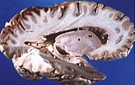
Sagittal section of the brain: gray matter outside and white matter inside.


Individual evidence
- ↑ David G. Myers: Psychology. Springer, 2005, ISBN 3-540-21358-9 , p. 479.
- ↑ Otto Grosser, arr. by Rolf Ortmann: Outline of the human development history. 6th edition. Springer, Berlin 1966, p. 78.
- ^ Hermann Voss , Robert Herrlinger : Taschenbuch der Anatomie. Volume III: nervous system, sensory system, skin system, increment system. 12th edition. Gustav-Fischer, Jena 1964; to chap. I. “The nervous system”, section “The internal structure of the spinal cord”, p. 8 and “The central cave gray” p. 48 f.