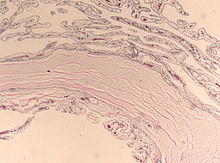
Plexus cyst
As plexus cysts ( choroid plexus cysts ) are harmless cystic structures without their own illness value in the range of the choroid plexus in the brain refers to an unborn child which are visible in one or both sides an oval or round shape. The size is usually 0.3 to 2 mm. Plexus cysts occur both unilocular (limited to one site) and multilocular (in multiple sites).
Basics
The inside of the human brain contains four chambers, the so-called brain ventricles . In these chambers, a specially shaped plexus of veins, the choroid plexus , forms the cerebrospinal fluid ( liquor cerebrospinalis ).
Cysts are small, fluid-filled cavities in tissue. As a rule, cysts are completely harmless and have no disease value of their own, that is, they are not dangerous or harmful to humans. They occur in the human body in or on various organs.
frequency
A plexus cyst is found in the unborn baby's brain in around 1 to 2% of all prenatal ultrasound examinations carried out in the second trimester of pregnancy (2nd trimester ) up to around the 28th week of pregnancy as part of prenatal diagnosis . Between the 16th and 20th week of pregnancy, minor cystic changes in the choroid plexus are more common in percentage terms than after the 20th week of pregnancy. Sometimes the posterior horn of a lateral ventricle is mistaken for a plexus cyst during an ultrasound scan.
meaning
Unless other peculiarities can also be determined, isolated plexus cysts (like other so-called soft markers ) do not have a disease value of their own, but are only statistically associated with a slightly increased probability of a chromosome peculiarity in the unborn child.
In the case of plexus cysts, this link refers specifically to Edwards syndrome (trisomy 18 or trisomy E), which occurs comparatively rarely with an average probability of 1: 3,000 to 1: 10,000. A plexus cyst can be found in the brain in about 43% of all unborn babies with trisomy 18.
As a rule, plexus cysts regress on their own and, according to current knowledge, have no influence on the health or the pre- and postnatal physical and cognitive development of the child. Only after the birth could it be observed in rare cases that plexus cysts in the third cerebral ventricle caused an obstruction of the circulation of the cerebral water , which resulted in the formation of internal hydrocephalus . Even smaller cysts in the area of the Foramina Monroi can result in bilateral but also unilateral hydrocephalus occlusivus. Corresponding prenatal observations have not yet been made on this rare phenomenon.
Procedure after the assessment
In the differential diagnosis , plexus cysts must be differentiated from subependymal cysts . These are often located at the transition from the core area in the endbrain ( nucleus caudatus ) to the diencephalon ( thalamus ) and are more often unilateral.
After a confirmed diagnosis of a plexus cyst in a baby's brain, the child's organs should be examined using fine ultrasound . An echocardiography of the heart is also advisable. In particular, if plexus cysts persist after the 28th week of pregnancy, if they are bilateral or particularly large, and especially if other physical signs can be found that often occur in unborn babies with Edwards syndrome , a chromosome examination after invasive diagnostics (e. B. chorionic villus sampling or amniocentesis ) should be considered after detailed advice and information on the advantages and disadvantages, the risks and the possible consequences.
An invasive prenatal diagnosis with subsequent chromosome analysis can diagnose or rule out trisomy 18 in the child with almost 100% certainty and the parents-to-be can have a pregnancy terminated or prepare for the birth of a special child if the result is positive .
literature
- Michael Entezami: Sonographic Malformation Diagnosis: Teaching Atlas of Fetal Ultrasound Examination. Georg Thieme Verlag, 2002, p. 222 ( online )
- A. Büttner and others: Colloid cysts of the third ventricle with fatal outcome: a report of two cases and review of the literature. In: International journal of legal medicine , 1997, pp. 260-266, PMID 9297582 .
- K. Hurt, O. Sottner, J. Záhumenský and others: Choroid plexus cysts and risk of trisomy 18. Modifications regarding maternal age and markers. In: Ceska Gynekol , 2007. pp. 49–52 (Czech)
- C. Papp, Z. Ban, Z. Szigeti, A. Csaba, A. Beke, Z. Papp: Role of second trimester sonography in detecting trisomy 18: a review of 70 cases . 2007, pp. 68-72, doi : 10.1002 / jcu.20290 .
- T. Dagklis, W. Plasencia, N. Maiz, L. Duarte, KH Nicolaides: Choroid plexus cyst, intracardiac echogenic focus, hyperechogenic bowel and hydronephrosis in screening for trisomy 21 at 11 + 0 to 13 + 6 weeks . In: Ultrasound Obstet Gynecol , 2007, pp. 132-135, PMID 18085527 .
- K. Demasio, J. Canterino, C. Ananth, C. Fernandez, J. Smulian, A. Vintzileos: Isolated choroid plexus cyst in low-risk women less than 35 years old. In: American Journal of Obstetrics and Gynecology 187 , 2002, pp. 1246-1249 PMID 12439513 .
Web links
- Jin Ho Jeon, Sang Weon Lee, Jun Kyeong Ko, and others: Neuroendoscopic removal of large choroid plexus cyst: a case report . 2005 (PDF; English)
- Plexus cysts on ultrasound (second trimester of pregnancy): [1] , [2]
- Course of a large singular (single) fetal plexus cyst
