Optic chiasm
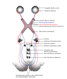
Optic chiasm (from Greek χίασμα chiasm "crossroads", from the Greek letter chi ( Χ ), and the Latinized form of the Greek οπτικόν OPTIKON "vision on"), and optic nerve ( s ) crossing , is the anatomical term for the intersection of the visual pathway where fibers of the optic nerve ( nervus opticus ) of one eye change sides and pull into the mutual optic cord tractus opticus .

In the optic chiasm, nerve fibers cross from the eye on one side to the opposite side. The extent of crossing fibers of retinal ganglion cells is in the individual vertebrates different. While in amphibians all fibers of an optic nerve are found in the contralateral optic tract, the proportion in primates such as humans is around 50%. With this half-crossing of the optic nerves ( semidecussatio nervorum opticorum ) only the fibers pull from the medial, nasal half of the retina to the opposite side and then in the tracus opticus to the contralateral hemisphere . The fibers from the lateral retinal halves of both eyes, which are located towards the temples (temporal), remain uncrossed and run in the optic tract to the ipsilateral hemisphere on the same side. This division is optimal for the almost completely binocularly captured field of view of a person.
Theories and theses on the evolution and function of the optic chiasm are dealt with under contralaterality of the forebrain .
Classification within the visual pathway

Light stimuli that were picked up by the photoreceptors of the retina and converted into a signal are processed neuronally within the layers of the retina. The neurites of the third afferent neurons in the ganglion cell layer leave the eye, crossing the retinal layers - like causing the blind spot - at the optic disc ( discus nervi optici ) and thus form the optic nerve of an eye, which extends from the eye socket through the optic canal into the canal Cranial cavity occurs. In humans, the optic nerve leading two together for about 3 million nerve fibers , each of a well of myelin sheaths enveloped Axon , the worked-in a special way signals from a particular area of the retina passes, each receptive field of his retinal ganglion cell.
Due to the imaging properties of the eye, sensory cells in retinal areas located towards the nose are stimulated by light emanating from objects that can be assigned to the temporal area of the visual field. The half of the retina, which is towards the middle and nasally, receives stimuli from the half of the field of vision of an eye, which lies towards the outside, temporally; With the temporal half of the retina it is the other way round; it is assigned stimuli that are in front of the nose in humans.
Animals with two eyes can absorb similar stimuli from an object with both of them if their eyes are positioned in such a way that the fields of vision that can be perceived by one eye (monocular) overlap. In humans, this is the case, which shapes their face, even without (convergent) eye movements, and the binocularly perceptible section of their field of vision is very large. Light stimuli from this area are in most cases imaged in such a way that those from the left binocular half of the visual field stimulate the nasal retina areas of the left eye and, at the same time, temporal retina areas of the right eye.
The signals obtained from the two different halves of the retina can be received and compared in the same brain region if they are brought together there. For this, the temporal can be led uncrossed to the same side (ipsilateral), while the nasal can be crossed; Then the brain region on the same side receives signals from corresponding network areas of both eyes, which correspond to an image of stimuli from the mutual (contralateral) half of the visual field. A comparison of the signal patterns created in one and the other eye enables spatially perceived vision.
The degree of crossover in the chiasm is quite different in vertebrates . In amphibians , 100% of the fibers usually cross. Almost all fibers cross in most birds , but only 60–70% in owls . In some predators about 75% of the fibers change, in many ungulates 90%. In primates like humans , about half of the optic nerve fibers cross, only the nasal ones, whereas in the chiasm those from the retinal periphery are below the macular ones .
Like any sensory information, apart from smelling, the optical information is first passed through the thalamus - more precisely the dorsal part of the corpus geniculatum laterale in the metathalamus - and switched over before it reaches the cerebral cortex, in this case the visual cortex , where it becomes more visual Information can be.
topography

The relationships between the optic chiasm (gray in the center of the picture) and the lamina terminalis , the III. Ventricles , vessels , as well as the pituitary gland (with adenohypophysis (light red) in front, neurohypophysis (dark red) behind) and the pituitary stalk ( infundibulum ).
The optic chiasm is a flat structure on the base of the brain, below which the anterior wall of the III. Ventricular- forming lamina terminalis . On each side there is an inner carotid artery , above it the anterior connecting artery , and at the rear it encompasses the infundibulum with the pituitary gland in a scissor-like manner . Because of the close proximity, tumors of the pituitary gland , for example, can lead to visual disorders, for example chiasma syndrome .
If the optic chiasm is severed in the median plane and with it the crossing fibers, the temporal halves of the visual field for each eye are omitted in humans, so-called bitemporal hemianopsia occurs , with severe restriction of the two-eyed visual field on both sides. A transection of the optic tract on one side, on the other hand, leads to a homonymous hemianopia in which the two halves of the field of vision of both eyes fall away.
literature
- Theodor Axenfeld (founder), Hans Pau (ed.): Textbook and atlas of ophthalmology. With the collaboration of Rudolf Sachsenweger and others 12th, completely revised edition. Gustav Fischer, Stuttgart et al. 1980, ISBN 3-437-00255-4 .
Individual evidence
- ↑ Franz-Viktor Salomon et al. (Ed.): Anatomy for veterinary medicine. Enke Stuttgart. 2nd ext. Edition 2008 ISBN 978-3-8304-1075-1 , p. 564.
- ^ Jeffery G: Architecture of the optic chiasm and the mechanisms that sculpt its development . In: Physiol. Rev. . 81, No. 4, October 2001, pp. 1393-414. PMID 11581492 .