Pillar valency

Under pillar value the utility of will teeth as abutment teeth tight or removable dental prosthesis understood. Abutment teeth are teeth that have to bear the load of replaced teeth. The pillar value is an expression of the tooth-related prognosis , against the background of a planned inclusion in various prosthetic restorations. The planned abutment teeth are to be evaluated for the planning of long-term durable dentures. The demarcation between usability and non-usability of teeth is difficult and often ambiguous. In everyday clinical practice, an appropriate risk assessment is essential when planning dentures . It is one of the most important dental skills and helps to reduce failures.
nomenclature
In addition to the term abutment tooth, there are other terms for teeth that are included in dental prosthesis treatments. This includes in addition to the bridge piers of the anchor tooth , the tooth pillar , the fixing element (in dentistry), the anchoring tooth (rather for construction elements in orthodontics) or the abutment tooth or clip tooth (for removable dentures for a tooth to which a bracket is attached to a bracket prosthesis ).
Basic value of the teeth
The number, length and diameter of the roots initially determine the basic value of a healthy abutment tooth. The basic value is divided into three classes:
1 = best value
2 = medium value
3 = restricted value
| top right | top left | |||||||||||||||
|---|---|---|---|---|---|---|---|---|---|---|---|---|---|---|---|---|
| 3 | 1 | 1 | 2 | 3 | 1 | 3 | 2 | 2 | 3 | 1 | 3 | 2 | 1 | 1 | 3 | Pillar valency |
| 18th | 17th | 16 | 15th | 14th | 13 | 12 | 11 | 21st | 22nd | 23 | 24 | 25th | 26th | 27 | 28 | Tooth- |
| 48 | 47 | 46 | 45 | 44 | 43 | 42 | 41 | 31 | 32 | 33 | 34 | 35 | 36 | 37 | 38 | designation |
| 3 | 1 | 1 | 2 | 2 | 1 | 3 | 3 | 3 | 3 | 1 | 2 | 2 | 1 | 1 | 3 | Pillar valency |
| bottom right | bottom left | |||||||||||||||
( Incisivi (incisors), canini (canines), premolars (small molars), molars (large molars)).
For example, the tooth roots of the lower incisors (32-42) are very thin and therefore cannot bear such high loads as the lower canines (33, 43), which have longer and thicker roots .
Load value of the teeth
The Basel university professor Gottlieb Vest has made his own classification of the pillar value and has designated this as the load value. The lowest value is 1 (lowest resilience), the highest value is 6 (greatest resilience). ( Dental schemes are written from the patient's point of view.)
| top right | top left | |||||||||||||||
|---|---|---|---|---|---|---|---|---|---|---|---|---|---|---|---|---|
| 4th | 6th | 6th | 4th | 4th | 5 | 3 | 4th | 4th | 3 | 5 | 4th | 4th | 6th | 6th | 4th | Exposure value |
| 18th | 17th | 16 | 15th | 14th | 13 | 12 | 11 | 21st | 22nd | 23 | 24 | 25th | 26th | 27 | 28 | Tooth- |
| 48 | 47 | 46 | 45 | 44 | 43 | 42 | 41 | 31 | 32 | 33 | 34 | 35 | 36 | 37 | 38 | designation |
| 4th | 6th | 6th | 4th | 4th | 5 | 2 | 1 | 1 | 2 | 5 | 4th | 4th | 6th | 6th | 4th | Exposure value |
| bottom right | bottom left | |||||||||||||||
( Incisors , canini , premolars , molars ).
According to Vest, the load value of the teeth to be replaced must correspond to the load value of the abutment teeth, i.e. at least equal to or greater than this, in order to guarantee a sufficient long-term prognosis for a bridge to be fabricated. For example, if the lower four front teeth 32-42, which have a load value of 2 + 1 + 1 + 2 = 6, are missing, the two lower canine teeth 33 and 43, which have a load value of 5 + 5 = 10, are sufficient as abutment teeth these teeth. If, however, the two molars 26 and 27 are missing (load value 6 + 6 = 12), according to Vest the two end teeth 25 and 28 are not sufficient as sole abutment teeth (load value 4 + 4 = 8). Therefore, tooth 24 should be included in a bridge denture (load value 4 + 4 + 4 = 12).
Criteria for the pillar value
In the second step, additional factors are checked with regard to the individual usability of a tooth.
Periodontal condition
The periodontal condition of a tooth largely determines the quality of the abutment. Periodontal diseases often make a tooth unusable due to the bone loss in the alveoli . Advances in periodontics have made periodontal renovation possible in many cases, which restores usability. Gum pocket depths of more than 6 mm reduce the pillar value considerably because the tooth is only anchored to a limited extent in the jawbone. According to Eduard Mühlreiter and Theodore Emile de Jonge-Cohen, the average root length is between 12 mm (lower front teeth) and 16 mm (upper canine teeth).
Surface of the periodontium
As periodontal ligament (periodontal membrane) is the connective tissue of parodontium (periodontal ligament), respectively. The Ante'sche law , set up in 1926 by the Canadian dentist Irwin H. Ante , calls for the total area of the periodontal ligament must correspond to the affected teeth of bone anchored roots of the abutment teeth at least the (theoretical) total of the periodontal ligament of the roots of. If this is not the case, the abutment teeth would be overloaded and further bone loss would result in the abutment teeth. Ante's statements, however, are not evidence-based and therefore not necessarily reliable. It is seen as a recommendation rather than a 'law' these days.
A simplified rule states that the number of abutment teeth must correspond to the number of teeth to be replaced. However, this simplified rule does not take into account any bone loss on the abutment teeth.
The Bonn university professor Søren Jepsen measured the average values of the root surfaces of the teeth in healthy periodontium. With these reference values, it is possible to calculate whether the sum of the root surfaces required by Ante's law corresponds to that of the teeth to be replaced. The individual case must be assessed on the basis of x-rays of the remaining teeth using the half-angle technique . Due to the many root variants of the wisdom teeth 18, 28, 38, 48, no average values are given for them.
| top right | top left | |||||||||||||||
|---|---|---|---|---|---|---|---|---|---|---|---|---|---|---|---|---|
| 431 | 433 | 220 | 234 | 273 | 179 | 204 | 204 | 179 | 273 | 234 | 220 | 433 | 431 | in mm² | ||
| 18th | 17th | 16 | 15th | 14th | 13 | 12 | 11 | 21st | 22nd | 23 | 24 | 25th | 26th | 27 | 28 | Tooth- |
| 48 | 47 | 46 | 45 | 44 | 43 | 42 | 41 | 31 | 32 | 33 | 34 | 35 | 36 | 37 | 38 | designation |
| 431 | 433 | 207 | 180 | 268 | 168 | 154 | 154 | 168 | 268 | 180 | 207 | 433 | 431 | in mm² | ||
| bottom right | bottom left | |||||||||||||||
( Incisors , canini , premolars , molars ).
Crown-root relation
By periodontal disease single or by overloading teeth (occlusal trauma), there is a degradation of the alveolar bone, in which the teeth are anchored. At the same time, the tooth roots can become visible through the simultaneous receding of the gums. As a rule of thumb, the length of the visible part of the tooth must not exceed the length of the root anchored in the bone, otherwise the leverage forces acting on the root would be too great, which could lead to tooth loosening.
Root shape
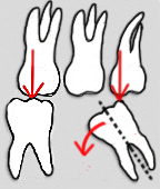
Teeth with splayed roots, as shown in the figure on the molars, have a favorable root shape . The pillar value is also increased by the shape of the individual root, which in the best case has a cylindrical shape (in the illustration the second tooth from the left - canine tooth 23). Unfavorable are conically tapered and short roots.
Degree of furcation
As a bifurcation (with two rooted teeth) or trifurcation (at three rooted teeth) the split-up point of the roots will be referred to in multi-rooted teeth. In a periodontally healthy tooth, they lie within the jawbone and are neither visible nor probable. Bifurcation and trifurcation are divided into four degrees of furcation. An exposed furcation caused by periodontal bone resorption creates a potential area of inflammation that is often difficult to clean. Depending on the severity, an exposed furcation can reduce the pillar value.
| Furcation degree 0 | Furcation not palpable |
| Degree of furcation 1 | Furcation entrance palpable |
| Degree of furcation 2 | Furcation clear, but not continuous, probable |
| Degree of furcation 3 | The furcation can be probed and is continuous on both sides |
Hemisected or premolarized teeth
A hemisection is the severing of a lower molar with a partial extraction of a tooth root. In the case of premolarization, the molar is also divided, but both roots are retained. This turns one molar into two premolars. Premolarization is a therapeutic measure used to remove an exposed bifurcation. The bifurcation creates an interdental space that is more accessible for cleaning. A hemisected or premolarized tooth has pillar value 3 only if the root length is complete and the remaining crown residue is high. In this case, the premolarized tooth parts are suitable for restoration with one or two crowns, but only to a limited extent as a supporting pillar for a bridge or removable denture.
Degree of tilt
Tilted teeth are not as resilient as straight teeth. The Sharpey fibers , on which the tooth is suspended in the alveolus (tooth socket), are stretched and loaded unevenly when subjected to stress. Tilting can create niches of dirt that can lead to inflammation. If it is tilted too much, it is difficult to prepare a common insertion direction for the denture. It can be overcome with a compensation attachment. Alternatively, the tooth can be straightened up again through orthodontic treatment. A tilt of up to 30 ° is tolerable. A greater tilt severely limits the usability. If there are no other factors that reduce the value, such teeth can be used as terminal abutment teeth. Teeth with a degree of tilt of more than 40 ° cannot be used as abutment teeth. Visually, a tilted tooth can appear to be straightened up by a crown, but the load always hits a tilted tooth.
Tooth mobility
Tooth mobility is measured in four degrees of relaxation (also degrees of mobility ), with four different classifications. Grade 0 and Grade 1 do not reduce the abutment value, Grade 2 requires comprehensive therapy of the tooth or only allows it to be used as a transitional restoration ( interim restoration ). At grade 3 there is no pillar valency. The measurements themselves can be carried out with the aid of a calibrated periodontal probe or electronically (Periotest).
Classification in statutory health insurance
| Loosening degree 0 | physiological mobility |
| Loosening degree I. | just palpable mobility |
| Loosening degree II | visible mobility |
| Loosening degree III | movable on lip or tongue pressure or axial mobility |
Tooth mobility is represented in the tooth status with Roman numerals .
Classification according to Carranza and Takai
| Mobility grade 0 | normal mobility |
| Mobility grade 1 | slightly more than normal agility |
| Mobility grade 2 | moderately more than normal mobility |
| Mobility grade 3 | strong mobility, faciooral or mesiolingual, combined with vertical mobility |
Classification according to Lindhe and Nymann
| Mobility grade 0 | normal mobility |
| Mobility grade 1 | horizontal mobility from 0.2 to 1 mm |
| Mobility grade 2 | horizontal mobility of 1 to 2 mm |
| Mobility grade 3 | horizontal mobility greater than 2.0 mm and / or axial mobility |
Knocking sound
The teeth can be checked for their knocking noise by tapping, for example by means of an instrument handle end. A bright knocking sound is evidence of a resonating bone in which the tooth is firmly anchored. The healthy Sharpey fiber apparatus couples the tooth well with the jawbone, and a dull knocking sound is a sign of reduced primary stability of the tooth. In this case, the periodontal gap is widened, which suggests reduced periodontal fixation of the tooth and thus reduced abutment value. The periodontal tissue is infiltrated by inflammation, and the coupling between tooth and bone is either absent or limited.
Endodontic condition
An irritant-free pulp (colloquially: "tooth nerve") is a prerequisite for a high pillar value of a tooth. Dentine is one of the most resistant organic materials. It consists of mineral nanoparticles and dental tubules that are embedded in a dense network of collagen fibers . The internal stresses in the nanostructure help to limit the formation and spread of cracks when exposed to stress. As the tiny collagen fibers shrink, the embedded mineral particles become increasingly compressed. The way of compression ensures that the innermost areas of the tooth are largely protected from cracks so that the sensitive pulp is not damaged.
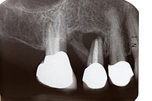
However, if the tooth is pulp (inflamed) or devitalized (dead), it must be treated endodontically in order to (also) achieve a corresponding pillar value. An endodontically treated tooth is more brittle and therefore more prone to breakage than a vital tooth. This can reduce the pillar value. After a root canal treatment, the root canal filling must extend to the physiological apex (root tip) and be at the edge. Periapical inflammation (in the bone in the area of the root tip) leads to the tooth becoming unusable as long as the inflammation has not healed or has been removed by a root tip resection (cutting of the root tip).
Carious destruction
The extent of carious destruction affects the usability of a tooth as an abutment tooth. If the clinical crown is almost or completely destroyed, it must be reconstructed using abutments, which in turn must be firmly anchored in the tooth roots. The abutments can be fastened using fillings with and without retention pins, using an adhesive attachment or using pin abutments . The diameter of a root post must be one third of the root diameter, the post length must at least correspond to the length of the tooth crown to be replaced. Only then is adequate retention of the post in the root canal ensured. However, posts weaken the tooth root, which reduces the value of the abutments. The value of the pillar depends on the type of structure; The decisive factor here is whether a post abutment cast from gold, a standardized Parapost titanium post with composite abutment , a glass fiber or carbon fiber post with composite abutment or a purely adhesively attached composite filling without a root post is used.
Ferrule effect
Teeth with a strongly widened canal entrance to the root canal and those without a barrel hoop preparation are to be assessed as critical, indeed as not sufficiently clinically resilient. The degree of destruction must allow adequate marginal fit of the artificial tooth crown. It is not enough if the artificial tooth crown is razor-sharp at the edge. The edge of the crown must firmly encircle the tooth in the form of a band with a width of around 2 mm ( ferrule effect ), otherwise the tooth is at risk of breaking. By Anthony W. Gargiulo et al. in 1961 the mean biological width was determined to be 2.04 mm. Of this, the periodontium takes up 1.07 mm and the marginal epithelium about 0.97 mm. If the tooth is destroyed to such an extent that this required width is not achieved, then - provided that the root length is sufficient - this ferrule area ("barrel hoop") can be created by means of a surgical crown extension . In the case of surgical crown lengthening, the edge of the bone around the tooth is removed until the remaining tooth is about 3 mm exposed, because the edge of the crown must not end directly at the bone boundary. A space for the formation of a gingival papilla in biological width must remain. Surgical crown lengthening, however, in turn shortens the root portion anchored in the jawbone, which in turn reduces the pillar value. The prognosis improves if a tooth has proximal contacts (contact with neighboring teeth), which can only be achieved on one side with terminal abutment teeth. Proximal contacts serve, among other things, for mutual support of teeth.
Retention form
The usability of a tooth and its pillar value include creating a retention form by grinding (preparation) the tooth. The retention of a crown on a tooth is not achieved by the fastening material alone. In addition, a slightly conical shape (5 ° to 8 ° cone angle ) must ensure retention of the tooth crown . The size of the retention area is also decisive for the hold of a crown. If a tooth is too badly damaged, or if it was already designed too conical in an earlier preparation, or if the crown stump is too short, the value of the abutments is considerably reduced. There is a risk, especially in the molar area, that the crown will detach from the tooth. The danger is particularly great in the lower jaw, since on the one hand the dentures are rigid and on the other hand the lower jaw body twists when the mouth is opened and under load. The attachment of the crown to the tooth must be able to permanently withstand this force difference. The contracted pterygoidei laterales muscles (outer wing muscles) compress the mandibular arch with the mandibular symphysis as a fixed point, which can deform the lower jaw by 0.1 to 1.0 mm.
Implants
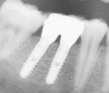
If there is sufficient bone for anchoring (circular ≥ 2 mm), after complete osseointegration (ossification), sufficient length (≥ 10 mm) and sufficient diameter (≥ 4 mm), the abutment value of implants corresponds to that of a healthy, natural canine (grade 1 ). Depending on which compromises have to be made in relation to the criteria mentioned, the pillar value of implants can decrease accordingly.
Milk teeth
To preserve a severely carious milk tooth , it can be reconstructed as a placeholder (for the pending eruption of the permanent tooth) with a simple, prefabricated crown that only remains for a few months to years until the tooth change . However, milk teeth are fundamentally unsuitable as abutment teeth because their roots are too weak. In addition, the milk tooth roots are resorbed during the tooth change. An exception can be a persistent deciduous molar if the permanent tooth is not positioned. If indicated, such a milk tooth can have an artificial crown. However, due to the short roots, it is not suitable for use as an abutment tooth.
Soft criteria for post usability
The soft criteria include those that do not in themselves change the pillar value. However, the usability of the pillar can be influenced by such additional factors.
Oral hygiene
It is possible that a tooth has a good abutment value, but poor oral hygiene on the part of the patient prevents its use, because the selected form of restoration then has little prospect of long-term success. For example, a tooth that has been severely damaged in terms of periodontics can be given a sufficient pillar value in a complex manner. However, if continuous aftercare and care are not guaranteed, the established pillar value is only a snapshot.
Planned dentures
The value of the pillar is also determined by which dental prosthesis is planned with which objective. A tooth can, for example, have sufficient abutment value for a transitional restoration (interim restoration). However, the same tooth can be unsuitable for long-term restoration. A tooth may also be part of a telescopic supply having sufficient valence pillar, because this at a tooth loss expandable is. The total supply by means of dentures would not be endangered by the loss of the tooth. However, the same tooth could no longer have sufficient abutment value for a fixed bridge restoration. If this abutment tooth were to be lost, the bridge restoration would be destroyed.
When planning a bridge or a partial denture , the statics and the forces that the abutment teeth will be exposed to must be determined. The abutment teeth are to be assessed as to whether they can withstand the expected loads, whereby a professional construction is assumed.
General illnesses
A generally increased risk of bone necrosis in the area of the alveolar process, for example after radiation therapy , chemotherapy or as a result of bisphosphonate medication , can reduce the pillar value.
Youthful teeth
In adolescents, the pulp cavity (tooth cavity) is wide. There is a risk of the pulp opening during preparation (grinding) of the teeth to accommodate a crown, which can result in limited usability as an abutment tooth. If necessary, a preparation that is gentle on the tooth substance, such as the Maryland bridge (adhesive bridge), can make a young tooth usable for a bridge restoration. The tooth is only prepared (ground) on the oral (inner) side. The tooth to be replaced is adhesively attached to the neighboring tooth with one or two wings . The Federal Joint Committee (G-BA) has expanded the guidelines for dental prosthesis supply in 2016: "For insured persons who have reached the age of 14 but not yet 21, the replacement of two incisors that are missing next to each other can be replaced by the Abutment teeth a single-span adhesive bridge with a metal frame with two wings or two single-span adhesive bridges with a metal frame with one wing each may be displayed. To replace an incisor, if there is sufficient oral enamel on one or both abutment teeth, a single-span adhesive bridge with a metal framework with one or two wings may be indicated. In the case of single-wing adhesive bridges to replace an incisor tooth, the tooth adjacent to the pontic of the adhesive bridge, which is not the support of a wing, should not need a crown and should not be provided with a crown in need of replacement ”.
Counter teeth
The load that a tooth has to bear also depends on the opposing teeth. If, for example, a restoration with a bridge is planned in one jaw and there is a partial or full denture in the opposing jaw , then the biting force is reduced. This means that the abutment teeth of the bridge have to absorb less load than in the case of opposing teeth with healthy teeth or implants. In this case, teeth with a reduced pillar value can also be used as bridge piers.
Patient wishes
If patients want dental restoration designs in which teeth with reduced abutment value are to be used, then prior information about the possible consequences is essential, which indicates the reduced length of stay of the denture. Time-consuming and costly treatments can be expected again in these cases after a shorter period of time. Section 630e of the German Civil Code (BGB), which was introduced by the law to improve patient rights in 2013, specifies the dentist's duty to provide information . The patient must be informed about all the essential circumstances for the consent, in particular about the type, scope, implementation, expected consequences and risks of the measure as well as its necessity, urgency, suitability and prospects of success with regard to the diagnosis or therapy. When providing information, reference should also be made to alternatives to the measure if several medically equally indicated and common methods can lead to significantly different burdens, risks or healing chances.
Economic efficiency requirement
In Germany, when drawing up a treatment and cost plan - taking into account the economic efficiency requirement of the statutory health insurance according to Section 12 of the Social Code Book V - the pillar value for the planned dental prosthesis is of decisive importance for obtaining a fixed allowance . If the prognosis of the tooth is questionable, the tooth falls out of the eligibility for subsidies.
literature
- Peter Pospiech: Pillar quality. In: Peter Pospiech: The prophylactically oriented supply with partial dentures. Thieme, Stuttgart et al. 2001, ISBN 3-13-126941-3 , p. 146 ff., ( Restricted preview . Accessed on February 8, 2017).
- Peter Pospiech: The prosthetic pillar. In: Military medicine and military pharmacy . Vol. 57, No. 2/3, 2013, pp. 63-66, ( digitized version ).
- Daniel Pagel: Prosthetics in periodontally damaged teeth. Risk assessment and therapeutic options. Spitta, Balingen 2014, ISBN 978-3-943996-34-0 (Excerpt: Online . Accessed February 8, 2017).
- Michael G. Newman, Henry Takei, Perry R. Klokkevold, Fermin A. Carranza: Carranza's Clinical Periodontology. 12th edition. Elsevier, St. Louis MO 2015, ISBN 978-0-323-18824-1 .
Web links
- S1 recommendation : fixed restorations for dental limited gaps , German Society of Oral and Maxillofacial Surgery . ( Digitized version ), August 1, 2012. Accessed February 11, 2017.
Individual evidence
- ^ MH Walter, Risk pillars value , Quintessenz Verlag, Berlin (2011). Retrieved July 30, 2015.
- ↑ Harald Schrenker: Compromises and limits in prosthetics . Spitta, 2003, ISBN 978-3-934211-61-2 , pp. 43-45 . Limited preview in Google Books . Retrieved February 9, 2017.
- ↑ Gottlieb Vest, Textbook of Dental Crown and Bridge Prosthetics: Volume 2, Bridge Prosthetics , Springer Heidelberg, New York 2013, ISBN 978-3-0348-7073-3 , pp. 101-102. Limited preview in Google Books . Retrieved February 9, 2017.
- ↑ a b P. Pospiech, Checklist prosthetic restoration . In: The prophylactic-oriented supply with partial prostheses , Thieme-Verlag (2001) ISBN 3-13-126941-3 . Limited preview in Google Books , pp. 144–149. Retrieved February 3, 2017.
- ↑ Eduard Mühlreiter (ed.), Theodore Emile de Jonge-Cohen, Anatomy of the human teeth , Arthur Felix Leipzig 1870.
- ↑ Irwin H. Ante, The fundamental principles of abutments, Michigan State Dental Society Bulletin 1926; Volume 8, pp. 14-23.
- ↑ M. Lulic, U. Brägger u. a .: Ante's (1926) law revisited: a systematic review on survival rates and complications of fixed dental prostheses (FDPs) on severely reduced periodontal tissue support. In: Clinical oral implants research. Volume 18 Suppl 3, June 2007, pp. 63-72, doi: 10.1111 / j.1600-0501.2007.01438.x , PMID 17594371 (review).
- ↑ German Dentists Calendar 2009 . Deutscher Ärzteverlag, 2008, ISBN 978-3-7691-3401-8 , p. 156.
- ^ G. Greenstein, JS Cavallaro: Importance of crown to root and crown to implant ratios. In: Dentistry today. Volume 30, Number 3, March 2011, pp. 61-2, 64, 66 passim, PMID 21485881 .
- ^ Søren Jepsen, Root Surface of Teeth . In: Bärbel Kahl-Nieke, Introduction to Orthodontics: Diagnostics, Treatment Planning, Therapy: with 10 tables . Deutscher Ärzteverlag 2010, ISBN 978-3-7691-3419-3 . Limited preview in Google Books , pp. 181–182. Retrieved February 8, 2017.
- ↑ E. Czochrowska, A. Stenvik, B. Bjercke, B. Zachrisson: Outcome of tooth transplantation: Survival and success rates from 17 to 41 years post-treatment . In: American Journal of Orthodontics and Dentofacial Orthopedics . 121, No. 2, 2002, pp. 110-119. Digitized
- ↑ Peter Kolling. Gerwalt Muhle, Compromises and Limits in Periodontology , Spitta Verlag Balingen 2003, ISBN 978-3-934211-62-9 , restricted preview in Google Books , pp. 101-107.
- ↑ Thomas Mayer, Compromises and Limits in Endodontology . Spitta Verlag 2005. ISBN 978-3-934211-84-1 , limited preview in Google Books , p. 80.
- ↑ Peter Eickholz, Glossary of Basic Terms for Practice: Periodontal Diagnostics Part 1: Clinical Plaque and Inflammation Parameters , In: Parodontologie 16 No. 1, Quintessenz Berlin et al. 2005, pp. 69–75.
- ↑ PAR guidelines of the Federal Dental Health Insurance Committee (PDF; 21 kB). Retrieved July 30, 2015.
- ^ FA Carranza, HH Takai, Clinical diagnosis, in Clinical Periodontology, Saunders, Elsevier, pp. 540-560 (2006). ISBN 0-323-18824-9
- ↑ M. Giargia, I. Ericsson, J. Lindhe, T. Berglundh, AM Neiderud: Tooth mobility and resolution of experimental periodontitis. An experimental study in the dog. In: Journal of Clinical Periodontology . Volume 21, Number 7, August 1994, pp. 457-464, ISSN 0303-6979 . PMID 7929857 .
- ↑ Peter Pospiech: The prophylactically oriented supply with partial prostheses , Georg Thieme 2002, ISBN 978-3-13-126941-6 , restricted preview in Google Books , pp. 150–152.
- Jump up ↑ Jean-Baptiste Forien, Claudia Fleck, Peter Cloetens, Georg Duda, Peter Fratzl, Emil Zolotoyabko, Paul Zaslansky: Compressive Residual Strains in Mineral Nanoparticles as a Possible Origin of Enhanced Crack Resistance in Human Tooth Dentin. In: Nano Letters. 15, 2015, p. 3729, doi: 10.1021 / acs.nanolett.5b00143 .
- ↑ Klaus M. Lehmann, Elmar Hellwig, Hans-Jürgen Wenz: Dental Propaedeutics: Introduction to Dentistry; with 32 tables . Deutscher Ärzteverlag, 2012, ISBN 978-3-7691-3434-6 , p. 46 . Limited preview in Google Books
- ↑ Norbert Schwenzer: Dental, Oral and Maxillofacial Medicine: General Surgery: 59 tables / ed. by Norbert Schwenzer and Michael Ehrenfeld. With contribution by Arzu Agildere… Georg Thieme, 2000, ISBN 978-3-13-593403-7 , p. 124 . Limited preview in Google Books
- ↑ A. Samran, S. El Bahra, M. Kern: The influence of substance loss and ferrule height on the fracture resistance of endodontically treated premolars. An in vitro study. In: Dental materials: official publication of the Academy of Dental Materials. Volume 29, number 12, December 2013, pp. 1280-1286, doi: 10.1016 / j.dental.2013.10.003 , PMID 24182949 .
- ^ HH Takei, RR Azzi, TJ Han: Preparation of the Periodontium for Restorative Dentistry . In: MG Newman, HH Takei, FA Carranza, Carranza's Clinical Periodontology , 9th Edition, Philadelphia: WB Saunders Company (2002).
- ↑ Anthony W. Gargiulo et al., Dimensions and relations of the dentogingival junction in humans . Journal of Clinical Periodontology (1961) Volume 32, pp. 261-267.
- ↑ F. Alpiste-Illueca: Morphology and dimensions of the dentogingival unit in the altered passive eruption , in: Medicina Oral Patologa Oral y Cirugia Bucal. 2012, p. E814, doi: 10.4317 / medoral.18044 .
- ↑ M. Nevins, HM Skurow: The intracrevicular restorative margin, the biologic width, and the maintenance of the gingival margin. In: The International journal of periodontics & restorative dentistry. Volume 4, Number 3, 1984, pp. 30-49, ISSN 0198-7569 . PMID 6381360 .
- ↑ U. Brägger, D. Lauchenauer, NP Lang: Surgical lengthening of the clinical crown. In: Journal of Clinical Periodontology . Volume 19, Number 1, January 1992, pp. 58-63, ISSN 0303-6979 . PMID 1732311 .
- ^ A. Padbury, R. Eber, HL Wang: Interactions between the gingiva and the margin of restorations. In: Journal of clinical periodontology. Volume 30, Number 5, May 2003, pp. 379-385, ISSN 0303-6979 . PMID 12716328 . (Review).
- ↑ Jan Hajtó, Retention and form of resistance in cemented crowns and bridges , ZMK 2010, digitized part 1, digitalized part 2 . Retrieved February 10, 2017.
- ↑ Harald Schrenker, Compromises and Limits in Prosthetics , Spitta Balingen 2003, ISBN 978-3-934211-61-2 . Limited preview in Google Books . Pp. 43-46. Retrieved February 8, 2017.
- ↑ K. Sivaraman, A. Chopra, SB Venkatesh: Clinical importance of median mandibular flexure in oral rehabilitation: a review. In: Journal of Oral Rehabilitation. 43, 2016, p. 215, doi: 10.1111 / joor.12361 .
- ↑ Peter Gängler, Thomas Hoffmann, Brita Willershausen, Conservative Dentistry and Periodontology: 66 Tables , Georg Thieme 2010, ISBN 978-3-13-593703-8 , restricted preview in Google Books , p. 177.
- ↑ Wolfgang Gernet, Reiner Biffar, Norbert Schwenzer, Michael Ehrenfeld: Dental prosthetics . Georg Thieme, 2011, ISBN 978-3-13-165124-2 , p. 110 . Limited preview in Google Books
- ↑ Hans H. Caesar: The training as a dental technician . Verlag Neuer Merkur GmbH, 1996, ISBN 978-3-929360-01-1 , p. 388 ff . Limited preview in Google Books
- ↑ S1 guideline, recommendation of the German Society for Dentistry, Oral and Maxillofacial Medicine (DGZMK) Fixed dentures for gaps delimited by teeth ( memento of the original dated February 4, 2017 in the Internet Archive ) Info: The archive link was inserted automatically and has not yet been checked. Please check the original and archive link according to the instructions and then remove this notice. , (PDF; 318 kB) 2012. Retrieved on February 4, 2017.
- ↑ Dental prosthesis guideline: Adaptation in Section D. II. Numbers 22 and 24 - Adhesive Bridge , Federal Joint Committee, entry into force on May 3, 2016. Accessed on February 7, 2017.
- ↑ Dominique Schaaf, Survival Time Analysis of Extension and Spanned Bridges - A Retrospective Longitudinal Study . Dissertation Justus Liebig University Gießen, VVB Laufersweiler Gießen 2011, digitized , p. 82. Accessed on February 7, 2017.


