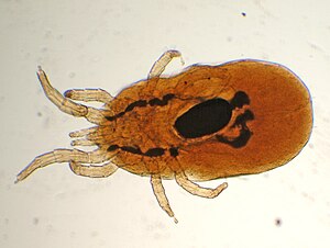
Red bird mite
| Red bird mite | ||||||||||||
|---|---|---|---|---|---|---|---|---|---|---|---|---|

Red bird mite under the microscope |
||||||||||||
| Systematics | ||||||||||||
|
||||||||||||
| Scientific name | ||||||||||||
| Dermanyssus gallinae | ||||||||||||
| De Geer , 1778 |
The red poultry mite ( Dermanyssus gallinae ) is a blood-sucking ectoparasite of birds. It affects wild birds such as songbirds as well as farm poultry, especially chickens , and also pet birds. As a false host , the red poultry mite also attacks mammals and humans, so-called bird-holder scabies.
features
Red mites are approximately 750 to 840 micrometers long and 400 micrometers wide (sexually mature females). They are soberly colored whitish gray. After a meal of blood, the red color of the blood shines through the intestines and the cover of the body (name); in advanced digestion this turns into brownish tones. As with almost all mites, the body is divided into two sections. The larger part of the trunk with the legs is called the idiosoma. At the front end, between the hips of the front legs, sits a smaller section that carries the mouthparts, the gnathosoma. In the genus Dermanyssus , the idiosoma is long-oval and broadly rounded at the back. It is predominantly softly sclerotic and flexible. There are more solidly embedded sclerotized plates called sclerites or shields. Dermanyssus has only one shield (dorsal shield) on the upper side. On the underside there are three smaller shields, the sternal shield, genital shield and anal shield (with the anus). The dorsal shield covers most of the upper side, it is elongated, broadly rounded at the front with clearly kinked front corners (or "shoulders"), narrowed behind long to the rear. The rear end is quite abrupt, almost straight, rounded in a truncated manner. The species can only be distinguished from other mite species of the same and related genera by the shape of the shields, but primarily by their bristling.
The mouthparts of Dermanyssus species are characteristically modified due to the parasitic way of life. The chelicerae are very elongated and bristle-shaped-cylindrical, especially their second limb is greatly elongated. The chela (scissors-shaped grasping forceps) at the tip has almost receded, it can only be seen in the electron microscope image. The chelicerae can be withdrawn into the trunk (far into the idiosoma) and stretched out when eating, they serve as piercing bristles to pierce the host's skin. The collapsed chelicerae form a food channel through which the blood is absorbed.
The males of the Dermanyssus species have an unpaired, central genital opening on the ventral side in front of the front edge of the ventral shield. Their chelicerae serve as mating organs ( gonopods ). They can also be recognized by the more heavily sclerotized body surface. So all three ventral shields are fused into one.
Life cycle
The species does not lay its eggs on the host, but in crevices within its nest or somewhere near it, in the case of animals in enclosures and cages in cracks and cavities of these. The red poultry mite hatches from the egg as a six-legged larval stage, goes through two eight-legged nymph stages after each molt, the last of which molts to become an adult. Nymphs can be distinguished from adults by their smaller, reduced shields. All stages are blood sucking. However, they do not remain on the host (like the Nordic bird mite, for example ) between blood meals, but leave it again immediately after the meal. They are therefore temporary ectoparasites, similar to e.g. B. the mosquitoes . Under favorable conditions (20 to 25 ° C, high humidity), the life cycle from the egg to the new oviposition of the females can be completed in one week.
Each oviposition, and each molt to the next stage, is preceded by a meal of blood. Three to four eggs are laid per egg-laying. During its lifespan, a female can produce around 300 eggs. The lifespan of a female reaches about 6 weeks at 25 ° C, it increases to 9 months at 5 ° C, but at this temperature neither growth nor development is possible. Animals without any opportunity to feed can survive for 34 weeks.
Ecology and way of life
Red mites are relatively less host-specific and are known from a large number of bird species (from eight orders), both kept by humans and those living in the wild. Economic problems exist in poultry breeding in particular, with all housing systems (cage, floor, free-range) being equally affected. The species is one of the economically most important pests in poultry farming, especially since it also transmits a number of infectious diseases. The species occurs worldwide, but economic damage is mainly known from Europe and, increasingly, South America, while it is less important in North America compared to the northern poultry mite.
The red poultry mite moves very quickly in relation to its own size. It attacks the birds only at night, during the day the parasite hides in the nest, in the cracks and crevices in the ceilings, walls, perches, etc. of the animals in the enclosure referred to as "gray mite"). At high densities and breeding birds, they can sometimes be found on animals during the day. The species can easily cover longer distances actively in search of a host and z. B. switch between enclosures and cages.
Red mites prefer temperatures between 20 and 30 ° C. At low temperatures (5 ° C) they survive and can even lay eggs, but these only develop further when temperatures rise. At temperatures well above 40 ° C, both the mites and their eggs die after a relatively short time. The animals survive, probably without any particular acclimatization, at temperatures around −10 ° C, but die quickly at −20 ° C (20 minutes). All stages of development are relatively sensitive to dehydration. They survived the longest in the experiment at 70% humidity.
Clinical picture
The harmful effect of the red poultry mite consists in sucking blood, triggering itching and inflammation and the associated stress of the infected animals. Chicks and young birds can die due to the constant blood collection even with moderate infestation. Direct deaths are also possible in breeding birds.
Sick birds scratch their plumage irritably. There is inflammation and long-lasting itching at the bite sites . The mite infestation is particularly visible on the legs of the birds. In extreme cases, the skin is very swollen, crusted and flaky. Individual skin areas gradually peel off.
The easiest way to detect the infestation is to put dead birds in white plastic bags or with "mite traps" (white adhesive tape) on the perches. You can also put a white cloth over the cage at night. If you find gray to blackish or red spots in the morning, this is a reliable indication of a mite infestation.
Economic damage
For poultry farmers, the economic damage caused by this parasite is particularly important, as infected animals are weakened and susceptible to other diseases because their immune systems are impaired. This also affects rearing, fattening and laying performance.
Combat
The animals are typically controlled with acaricides in powder form ( carbamates , pyrethroids , pyrethrum ). Ivermectin has proven to be very effective . Fluralaner has also been approved for use in drinking water since 2017 .
The removal of the mites from stables is more problematic. Here all hiding places must be thoroughly cleaned and treated with acaricides. Alternatively, a 2-component disinfectant based on peroxyacetic acid and hydrogen peroxide can be used.
An alternative to acaricides are silicate dust ( kieselguhr ). The mode of action is based on a drying effect on contact. Another possibility is to coat the underside of the perches with vegetable oil (basically all oils). In doing so, the oil clogs the pores and suffocates all stages of the mites.
A natural-based repellent can be used in laying farms to add soaking water . This does not cause the mites to die off, but prevents the mites from sucking blood and thus interrupts the reproductive cycle.
Double-sided tape on the ends of the perches can prevent the mites from migrating from the hiding places to the chickens and back.
Human infestation
| Classification according to ICD-10 | |
|---|---|
| B88.0 | Other acarinosis [mite infestation] |
| ICD-10 online (WHO version 2019) | |
Red mites normally only feed on the blood of bird species and can only complete their life cycle with this. If hungry mites but no birds are available, they try to suck blood from all warm-blooded organisms, including humans. Infestation shows up as unspecific arthropod dermatitis with red wheals (papules) with blistering and severe itching. The sting itself usually goes unnoticed, only the itching that sets in after a few hours draws attention to the attack. The hollows of the knees, the bends of the elbows and the navel region are preferred. Since the mites leave humans immediately after the act of sucking and are rarely noticed during this time, they are hardly ever found directly, which can often lead to misdiagnosis.
The infestation is widespread as bird owner dermatitis, especially among poultry breeders and keepers or pigeon breeders. But it can also be based on city pigeons nesting wildly on buildings . There is a particular danger here if the pigeons had reached high densities, but then suddenly disappeared, for example as a result of a fight. Roof apartments are often affected; in the examples mentioned, it was hospitals. The clinical picture, called gamasoidosis, remains local and allergic reactions are not reported. The transmission of bacteria or viruses to humans is considered possible in principle, but has also not been proven.
Taxonomy
The genus Dermanyssus comprises a good 20 species, of which only Dermanyssus gallinae (in the broader sense) occurs in bird species kept by humans. The other species in the genus are usually much more host-specific. However, Dermanyssus hirudinis , which is also widespread in Europe, has a similarly wide host range (songbirds, swallows, pigeons, ducks, owls ...), but it never occurs on chickens. In molecular studies (comparison of DNA sequences), the species gallinae was found to be well differentiated from the other species of the genus described. However, it then consists of several genetically separate but morphologically indistinguishable lines of development.
Web links
Individual evidence
- ↑ a b Birgit Habedank: The tropical rat mite Ornithonyssus bacoti and other predatory mites - rare parasites of humans in Central Europe. In: Horst Aspöck (Wiss. Red.): Amoebas, tapeworms, ticks ... Parasites and parasitic diseases of humans in Central Europe (= Denisia. 6 = Catalogs of the Upper Austrian State Museum. NF No. 184). Upper Austrian State Museum, Linz 2002, ISBN 3-85474-088-3 , pp. 447-460, ( digitized version (PDF; 1.58 MB) ).
- ↑ a b Antonella Di Palma, Annunziata Giangaspero, Maria Assunta Cafiero, Giacinto S. Germinara: A gallery of the key characters to ease identification of Dermanyssus gallinae (Acari: Gamasida: Dermanyssidae) and allow differentiation from Ornithonyssus sylviarum (Acari: Gamasidae: Macronyssidae. Macronyssidae ). In: Parasites & Vectors. 5, 2012, pp. 104-114, doi : 10.1186 / 1756-3305-5-104 .
- ^ William A. Phillis III: Ultrastructure of the chelicerae of Dermanyssus prognephilus Ewing (Acari: Dermanyssidae). In: International Journal of Acarology. Vol. 32, No. 1, 2006, pp. 85-91, doi : 10.1080 / 01647950608684446 .
- ↑ a b c Helena Nordenfors, Johan Hoglund, Arvid Uggla: Effects of Temperature and Humidity on Oviposition, Molting, and Longevity of Dermanyssus gallinae (Acari: Dermanyssidae). In: Journal of Medical Entomology. Vol. 36, No. 1, 1999, pp. 68-72, doi : 10.1093 / jmedent / 36.1.68 .
- ↑ A. Kirkwood: Longevity of the mites Dermanyssus gallinae and Liponyssus sylviarum. In: Experimental Parasitology. Vol. 14, No. 3, 1963, pp. 358-366, doi : 10.1016 / 0014-4894 (63) 90043-2 .
- ↑ a b Lise Roy, Ashley PG Dowling, Claude M. Chauve, Thierry Buronfoss: Delimiting species boundaries within Dermanyssus Dugès, 1834 (Acari: Dermanyssidae) using a total evidence approach. In: Molecular Phylogenetics and Evolution. Vol. 50, No. 3, 2009, pp. 446-470, doi : 10.1016 / j.ympev.2008.11.012 .
- ↑ Alphabetical directory for the ICD-10-WHO version 2019, volume 3. German Institute for Medical Documentation and Information (DIMDI), Cologne, 2019, p. 180
- ↑ Pierre Auger, Jacques Nantel, Nicole Meunier, Robert J. Harrison, Robert Loiselle, Theresa Gyorkos: Skin acariasis caused by Dermanyssus gallinae (de Geer): an in-hospital outbreak. In: Canadian Medical Association Journal. Vol. 120, No. 6, 1979, pp. 700-703, PMC 1819175 (free full text).
- ^ Anne P. Bellanger, Christian Bories, Françoise Foulet, Stephane Bretagne, Françoise Botterel: Nosocomial Dermatitis Caused by Dermanyssus gallinae. In: Infection Control & Hospital Epidemiology . Vol. 29, No. 3, 2008, pp. 282-283, doi : 10.1086 / 528815 .
- ↑ A. Kavallari, T. Küster, E. Papadopoulos, LS Hondema, Ø. Øines, J. Skov, O. Sparagano, E. Tiligada (2018): Avian mite dermatitis: Diagnostic challenges and unmet needs. Parasite Immunology 2018: 40: e12539. doi: 10.1111 / pim.12539
- ^ Lise Roy, Claude M. Chauve: Historical review of the genus Dermanyssus Dugès, 1834 (Acari, Mesostigmata: Dermanyssidae). In: Parasite. Vol. 14, No. 2, 2007, pp. 87-100, doi : 10.1051 / parasite / 2007142087 .
- ↑ Lise Roy, Ashley PG Dowling, Claude M. Chauve, Thierry Buronfoss: Diversity of Phylogenetic Information According to the Locus and the Taxonomic Level: An Example from a Parasitic Mesostigmatid Mite Genus. In: International Journal of Molecular Sciences. Vol. 11, No. 4, 2010, pp. 1704-1734, doi : 10.3390 / ijms11041704 .