Silk secretion
Silk secretion involves the production of silk components and their secretion by an animal . The silk production of the silkworms is known . The same fine-grained material can by most larvae of insects , some of arachnids and some shells by silk glands or by an epithelium excreted be. Depending on the degree of hardening, silk is a biological cement substance or a tough elastic fiber . It consists to a large extent of biopolymer structural proteins . The mother-of-pearl of the mussel shells also contains silk components.

Homologous and analog developments
The first facilities for silk production are to be found in the common ancestors of molluscs and articulated animals. In the course of further evolution , this resulted in a heterogeneity of solutions that has not yet been fully clarified.
While the essential silk component, the silk protein with several polar repeat sequences and in the β-sheet conformation, is presumably homologous , the silk-producing tissues from original ectodermal systems have developed in parallel several times . This also applies to the class of insects .
On the basis of the various anatomical conditions, the molecular structure of the silk components and the relationships of the insect groups, 23 different paths for the evolution of silk production in insects were determined.
Silk-producing animals
Some animal names refer directly to the use of endogenously produced threads with name elements such as web , silk , spinning or weaving :
- Web spiders
- Orb web spider
- Spinner (several butterfly families)
- be crazy
- Spider mites
- Silk spiders
- Silkworms
- Weaver ants
- Weaving spiders
But: Harvesters have no silk or spinneret glands, but stink glands . Weaver birds also do not produce threads, but rather artfully intertwine found nesting material.
Two recent groups of animals can either produce silk or silk components, or they have organ parts that are homologous to the silk glands : members of the molluscs ( phylum Mollusca) and arthropods (phylum Arthropoda).
Molluscs
Many shell molluscs (sub-tribe Conchifera), including some cephalopods (such as nacelles ), form shells whose innermost layer (the hypostracum) consists essentially of mother-of-pearl , which contains silk proteins.
Mussels (class Bivalvia, e.g. mussels ) produce mussel silk ( byssus ) to attach, often only in the juvenile stage. Byssus is homologous in structure to the silk of insects; it contains the silk proteins fibroin and sericin.
arthropod
Most classes of arthropods have numerous members who are capable of producing silk or the silk proteins fibroin and sericin, at least in certain age forms. The expression of silk proteins is not known from some , but organ structures that are homologous to silk glands can often be recognized:
| Arthropod (arthropoda) |
|
|||||||||||||||
|
|
Millipede
In the class of millipedes (Myriapoda), many species produce silk-like secretions that harden outside the body, e.g. B. bipedes .
Crustaceans
A production of silk components by crustaceans (Crustacea) is hardly known.
Some amphipods (amphipods), for example. B. Crassicorophium bonellii have silk glands on their feet.

Most sessile barnacles form a type of cement to attach themselves to. This secretion is homologous to silk secretions.
insects
Many insect larvae can produce silk, mostly to protect themselves in a cocoon during pupation .
Claw bearers
Among the arachnids only members of the order of the spider spider , spider mite and pseudoscorpion can produce silk and use it to form silk threads, often to make dwellings or fishing gear. No silk secretions are known from other orders of arachnids (e.g. harvestmen). Nine families of spider mites (prostigmata, acariformes) are known to be able to produce silk, e.g. B. Tetranychus .
Of horseshoe crabs (Limulidae) no silk products are known. However, there are anatomical homologies of their opisthosomal segments 4 and 5 with the spinnerets of the arachnids.
Anatomical structures for silk secretion
The silk-producing tissues have developed in parallel several times from the original ectodermal systems .
The two oldest spinneret fossil finds come from the middle Devonian and are 386 and 374 million years old, respectively. They are the earliest evidence of silk-producing animals. These spinning glands are attributed to the Attercopus fimbriunguis spider .
Not all organs of all silk-producing animals have been examined in more detail. Therefore, only a few well-known anatomical structures are shown as examples:
In shell molluscs
Shellfish of various orders secrete the silk components of mother-of-pearl through their epithelium.
In byssus-producing mussels, silk glands are located in their feet.
With amphipods
Marine amphipods ( Crassicorophium bonellii ) have silk glands on their feet that give off filamentous secretions.
With insects
The known spinneret glands of most insects (larvae) contain similar cells in the ultrastructure and consist of a mostly multi-wound tube for the production of the silk components, the rear end of which secretes the liquid-sticky silk through a spinneret or nozzle. The thread hardens quickly after it emerges, even with aquatic forms.
The silk-producing tissues of the insects developed several times in parallel from homologous ectodermal systems . They emerged from at least four different starting tissues in the course of phylogenesis :
- Epidermal cells in connection with the bristle organ
- Accessory glands of the genital organs
- Malpighian vessels of the larvae
- Labial glands of the larvae of Amphiesmenoptera (i.e. butterflies and caddis flies )
The caterpillars of butterflies and the larvae of caddisflies can Labialdrüse to silk glands to be converted, with which they can spin silk threads. The silkworm caterpillar arranges the silk thread around itself by counteracting it with rhythmic head movements.
The larvae of some hymenoptera also produce silk with their labial glands. It is unclear whether these originated homologously to the Amphiesmenoptera or in parallel.
Opisthosomal glands of the spider

The common ancestors of the web spiders had 4 pairs of silk glands on their 4th and 5th opisthosomal segments. While such opisthosomal attachments are absent in recent spiders, homologous structures are present in horseshoe crabs ( Limulus ).
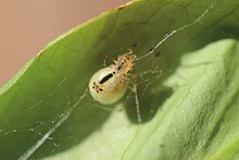
In the silk-producing spiders there are 4 to 6 silk glands or spinneret glands (2 or 3 pairs each) on the underside of the abdomen in the fourth and fifth abdomen segment. You can provide different types of silk for different tasks (glue threads, cocoon, shackle threads). These glands are externally recognizable as thickened spinnerets . The spinnerets are extremely mobile on the well-agile abdomen, with the muscles causing flexion and hemolymph pressure stretching. The spinnerets can also be spread apart to form “silk dabs” as anchoring points for the thread.
Many spiders can store differently designed spinning bobbins in front of their spinning glands when the silk thread emerges. These are tiny openings made of fine tubes (some cribellate spiders only 10 nm in diameter). A spinneret can be equipped with several different spinning bobbins for the production of threads of different thicknesses for different requirements (e.g. for abseiling threads, adhesive threads, alarm threads, cocoons).
The spinning plate of some trapped wool weavers (a plate-like converted spinneret, called cribellum ) can hold up to 50,000 spinning bobbins. Many cribellate spiders have a comb-like calamistrum on the metatarsus (last leg link) of the fourth pair of legs, with which their very fine silk wool can be combed. These anatomical structures (cribellum and calamistrum) only occur in cribellate spiders.
In addition to their spinning glands, Ecribellate spiders have glue-producing glands with which threads are glued in, which then serve as particularly sticky catch threads.
Adhesive secretion on the feet of tarantulas
In tarantulas of the genus Aphonopelma , additional spinning glands on their tarsi have been reported, which improved adhesion . However, this is contradicted by the fact that this observation is wrong and that the alleged spinning bobbins are actually chemosensors . The publications are controversial.
Spinning apparatus of the pseudoscorpions
Pseudoscorpions have spinnerets in their chelicerae (mouthparts).
Physiology of silk secretion
Provision of the soluble components
In the silk glands, the components of the silk are provided in an aqueous salt solution . The silk components are present in the gland as an aqueous solution until the secretion is squeezed out.
So that the areas of the silk proteins responsible for the firm cross-links do not clump together in the silk glands, they are first synthesized as precursors , which can be kept in the aqueous form. "Regulation areas" at the C-terminal end of the protein molecule cover those areas which could come into contact with one another in order to then form cross-links. Polar areas of the molecule are facing outwards, lipophilic areas facing inwards. This ensures good solubility in an aqueous medium.
Pressing out and consolidation
When squeezed out through the spinneret orifices, salt is retained and the silk proteins are secreted as a thin, sticky, liquid thread . As a result of the reduction in the salt concentration, the silk proteins can attach to one another and build up cross-links; solid silk fibrils are formed from these molecular associations. Depending on the function, remaining salts can leave the thread sticky for a longer period of time or, if the salt residue is very low, it can harden quickly. The outlet opening (bobbin) determines the thread thickness.
When the proteins emerge from the silk gland, they find a significantly reduced salt concentration and altered composition. The two ionic bonds of the regulator areas become unstable, the molecules change their folding and the molecular contact areas are exposed. The flow in the narrow spinning channel also creates strong shear forces, which causes the molecules to approach each other. The long protein chains are aligned parallel to one another based on their polar clusters. Now the areas responsible for cross-linking are right next to each other, creating a stable silk thread. Due to the special molecular arrangement of the amino acids involved, the threads produced are very tensile and at the same time highly elastic.
Molecular structure of silk
Insect and spider silk
The main components of the silk from silk spinners are the two structural proteins fibroin and sericin , which occur in the silk of the silk spinner in a ratio of 7: 3, without larger proportions of other components. The particularly stable properties of silk are primarily explained by the molecular structure of the silk protein fibroin, which makes up the main part of silk. It is a long-chain fiber protein (a β- keratin ). Sericin forms a matrix in which the long fibroin molecules can move and stick together shortly after they have been squeezed out. The related proteins spidroin 1 and spidroin 2 exist in spiders .
State of research
Insect and spider silk from only a few organisms have so far been investigated and only a few components have been molecularly characterized. Some processes can only be described for a single organism. The meager findings must therefore usually represent the silk production of other organisms that has not yet been analyzed in detail.
| Silk component | organism | publication |
|---|---|---|
| Fibroin | Silk moth ( Bombyx mori ) | numerous investigations |
| Sericin | Silkworms | |
| FLAG protein | Silk spiders ( Nephila ) | |
| sp160 protein | Mosquitoes | |
| sp185 protein | Mosquitoes | |
| sp220 protein | Mosquitoes |
The arrangement of the molecular components of the spider silk is also examined by means of X-ray structure analyzes. They show that spider silk consists of ordered (crystalline) and disordered (amorphous) areas. The crystallites cause the scattering contributions (Bragg peaks) on the detector. Disordered areas can be seen as a ring-shaped scattered background (amorphous halo). The evaluation of such an X-ray diffraction pattern makes it possible - among other things with the help of Miller indices - to determine the shape, extent and orientation of the crystallites.
Structure of the fibroin
The structure of the silk protein fibroin results from the multiple folding and alignment in four structural levels.
- The primary structure of fibroin is several polar repeats . The predominantly repeating sequence of amino acids in insect fibroin is Gly - Ser -Gly- Ala -Gly-Ala. The amino acid sequence of the spider fibroin has been described with poly alanine clusters 8-10 alanine residues next to glycine rich clusters of 24-35 amino acid residues.
- Fibroin is a β- keratin . The predominant secondary structure in the silk thread is the anti-parallel β-sheet . These stacks of sheets of several clusters form crystalline molecular regions.
- The tertiary structure of fibroin consists of two identical subunits, which are parallel to each other, but facing opposite. This arrangement is stabilized by hydrogen bonds and hydrophobic interactions between the subunits.
- In the quaternary structure , crystalline areas of fibroin molecules aligned parallel to one another are stabilized in their position to one another by intermolecular hydrogen bonds . Hydrophobic interactions between the fiber proteins also contribute to the stabilization of the silk thread.
Fibroin of the silk moth can exist in at least three conformations , from which different qualities of the silk thread result: silk I, II and III. Silk I is the natural state of the thread, silk II is found in the spooled silk thread. Silk III forms in an aqueous state at interfaces .
shine
The fibroin molecules arrange themselves in parallel in the silk thread when they exit the silk gland (paired β-sheets). Fixed cross-links develop between the areas provided for this purpose. The shine of the silk is based on the reflection of the light on these multiple molecular parallel layers. This optically active structure is not made responsible for the iridescent sheen of the mother-of-pearl, but rather its layered fine structure.
Additives
In addition to fiber proteins, insect silk also contains soluble (soluble in propylene glycol or glycerine) scleroproteins and other components:
Spider silk often contains antimicrobial components for the design of dwellings. Pheromones are often added to the silk , which allows species or gender to be identified. Catch threads are often equipped with adhesive additives or glue droplets. Salts, which adsorb humidity, are used to keep silk or adhesive substances moist.
Shell silk
Due to its origin in bivalve mollusks, byssus has a different structure than silks made from arthropods . Byssus contains at least nine characterizable proteins.
Silk of the amphipods
The silky threads of the amphipods consist of mucopolysaccharides and proteins. These have a high degree of β-sheet secondary structures with a clear content of polar amino acids.
Cement of the barnacles
The silk components of the cement of the barnacles (e.g. Megabalanus rosa ) contain insoluble proteins, rich in polar amino acids. From this, the fibroin-like protein Mrcp-20k (M. rosa cement protein with a molar mass of 20 kDa) was characterized, which is composed of six repeat clusters of around 30 amino acids each. The coding gene contains 902 base pairs, the precursor protein consists of 202 amino acids (20,357 Da), including a cysteine-rich signal sequence of 19 amino acids. However, the functional cement protein does not contain cysteine or disulfide bonds.
Functions of silk secretion
Silk is a highly resilient material with a very low weight. It is four times more resilient than steel and can be stretched three times in length without tearing.
In mollusks
Mussels use the silk secretion ( called byssus ) to attach themselves to solid ground with adhesive threads, especially in the surf zone . Fig clams spin nets from byssus threads and can use them to fix solid objects.
With many articulated animals
Egg cocoon
Spiders and many other articulated animals cover their eggs with fine silk to form an egg cocoon . Usually a stronger silk serves as external protection. Female wolf spiders carry their egg cocoons with them when hunting.
Rappelling
Many tensioners and most weaving spiders can abseil themselves on their silk thread. When climbing, many spiders pull a thread behind them, which is used to abseil when falling. For shedding, most spiders also use a spider thread to abseil out of the old skin (see also: Spiders # growth and molting ).
Flying thread
Spiders (Araneae), spider mites (Tetranychidae) and the larvae of many moths (Lepidoptera) produce a thread of flight ( English ballooning ) on which z. B. let the young spiders carry away in the wind in late summer (Indian summer) (aerial plankton ). Charles Darwin reported in his diary in 1832 that 100 km off the coast of South America countless small spiders got caught in the rigging of his research vessel.
In insect larvae
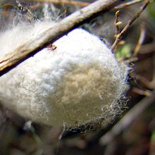
Many insects produce silk as larvae in order to create a cocoon as a protective cover for their pupation . The cocoon of the silk moth consists of a single thread up to 900 m long.
Cockroaches and Mantis (collectively sometimes referred to as Oothecaria) put their packages in fixed-curing cocoons ( oothecae ) from where the eggs are inseparably bonded together. Oothecae are quite resistant to predators and to mechanical or chemical influences.
There are also other functions of silk secretion:
- Butterfly caterpillars of many tooth moths and plant wasp larvae , e.g. B. Sawfly wasps , together form a web of silk threads in order to protect themselves from predators (e.g. songbirds). Some species from these groups build elaborate protective nests out of silk, which serve as hiding places for rest periods (e.g. processionary moths ).
- Aquatic larvae of the caddis flies stick together with their silk secretion species-specific substrate particles (sand, stones or plant material) to form a quiver that serves as a transportable body shell, which they also use for pupation. Other caddis flies build drift nets or catch nets similar to spider webs across the water current to catch food particles. Others weave security threads to prevent them from drifting themselves.
- Weaver ants use the secretion of their larvae to sew together leaves to form a nest .
With weaving spiders
Spermatophores
Like many other arthropods , male spiders often use larger packets of seeds ( spermatophores ) to transmit to the female because they lack a penis. Spider silk is usually used to cover the spermatophores.
Many male spiders produce parcels of sperm bundled in silk.
Sticky fishing gear
Many spiders can tie up their prey with sticky silk.
Many spiders also make fishing gear from spider silk.
Wheel nets are composed of different types of thread of different strengths and adhesive strength. Stable, non-adhesive threads form the basic network structure and are laid out first during construction. A non-adhesive auxiliary spiral defines the structure. The tightly drawn catch spiral made of adhesive threads is attached to this.
Tremble spiders and hood-web spiders build irregular space nets from which hanging threads covered with glue droplets hang.
Ecribellate spiders use threads covered in glue.
Non-stick fishing gear
Agelenidae lurking in a silk-lined hoppers for prey.
Trap-door spiders and link spiders lurk in their apartment tubes for touch signals from their alarm and trip threads.
Wallpaper spiders build a well-camouflaged catch hose and lurk in their living tube.
The fine trapping wool of the cribellate spiders has no adhesive strength, but is used to create very effective traps : The basic construction consists of strong axis threads onto which the extremely fine trapping wool is brushed, in which prey animals become hopelessly entangled.
Cast web spiders use cast webs. Some spiders (e.g. Dinopsis ) only have tiny webs to catch their prey, like a landing net.
Residential buildings
Many spiders line their caves with their silk or use it to build free webs of living space.

Water-dwelling water spiders hold a supply of air with their underwater webs, which they previously transported in portions in their body bristles. Consumed oxygen in the air bubble is regenerated from the water, because the air bubble functions as a "physical gill" or plastron . Conversely, the plastron releases carbon dioxide into the water.
Silk for communication
With some orb web spiders, males willing to mate are only allowed to approach after they have triggered a specific vibration via plucking signals. Jumping spiders and Lycosoidea also use silk and its vibrations for courtship and communication.
With pseudoscorpions
Pseudoscorpions create web nests of 5 to 7 mm in size for periods of rest and for wintering. To lay eggs, female pseudoscorpions create webs or special multi-chambered brood chambers.
With spider mites
Spider mites use a web in order to inhabit the underside of plants, e.g. B. Tetranychus , or to let the wind carry you. Cunixidae and Bdellidae produce webs for laying eggs and form silk nets, sometimes also for their spermatophores or for markings in intra-species communication.
Use by humans
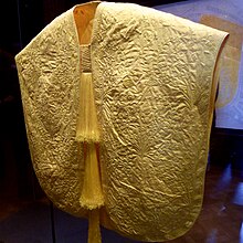
People use or used silk from the mulberry spinner, tussa, weaving spider (e.g. the garden spider ) or the noble pen shell ( Pinna nobilis L.), which lives in the Mediterranean , in order to obtain silk yarns by spinning , for the production of textile fabrics, ropes or bullet-resistant Dress.
The keeping of the silkworms and the extraction of the raw silk is called sericulture and has been practiced for 4800 years. It produces around 95% of all natural silk.
Woven fabrics made from shell silk or pen shell wool are known as byssus , but the term is also used for linen and byssos for cotton fabrics. Byssos fabric is finer than silk, because of its durability and the complex extraction it was very valuable and especially in the Middle Ages very popular among high dignitaries.
Fossil evidence
Fossil finds of silk products are rare. However, fossilization experiments with silk showed that tuff formation is possible through silk from aquatic insect larvae , especially in winter from caddis flies and in summer from black flies .
In amber , silk threads, also with adhering droplets of glue or trapped prey, were found from spider webs from the early Cretaceous period (around 130, 110 and 40 million years old).
Individual evidence
- ↑ a b c d e f Tara D. Sutherland, et al. : Insect silk: one name, many materials . In: Annual review of entomology . 55, September 3, 2009, pp. 171-188. doi : 10.1146 / annurev-ento-112408-085401 .
- ↑ a b c d e f František Sehnal, Hiromu Akai: Insect silk glands: their types, development and function, and effects of environmental factors and morphogenetic hormones on them . In: International Journal of Insect Morphology and Embryology . 19, No. 2, 1990, pp. 79-132. doi : 10.1016 / 0020-7322 (90) 90022-H .
- ↑ a b Katharina Gries: Investigations of the formation processes and the structure of the mother-of-pearl of abalons. Dissertation, University of Bremen, 2011 ( PDF ; 25.4 MB)
- ↑ Katharina Gries: Electron microscopic examinations of the polymer / mineral composite material mother-of-pearl. Diploma thesis, University of Bremen, 2007 - ( Memento of the original from December 26, 2013 in the Internet Archive ) Info: The archive link was inserted automatically and has not yet been checked. Please check the original and archive link according to the instructions and then remove this notice. (PDF; 4.1 MB)
- ↑ www.diplopoda.de For the egg-laying of the diplopods
- ↑ a b c Katrin Kronenberger, Cedric Dicko and Fritz Vollrath: A novel marine silk . In: Natural Sciences . 2012, pp. 1–8. doi : 10.1007 / s00114-0853-5 .
- ↑ a b Kei Kamino: Novel barnacle underwater adhesive protein is a charged amino acid-rich protein constituted by a Cys-rich repetitive sequence . In: Biochemical Journal . 356, No. Pt 2, 2001, p. 503. PMC 1221862 (free full text).
- ↑ a b c d Jacqueline Kovoor: Comparative structure and histochemistry of silk-producing organs in arachnids . In: Ecophysiology of spiders . Springer, Berlin Heidelberg 1987, IV, p. 160–186 (English, springer.com [accessed March 16, 2013]).
- ↑ a b c d P. A. Selden, WA Shear, MD Sutton: Fossil evidence for the origin of spider spinnerets, and a proposed arachnid order. In: Proceedings of the National Academy of Sciences . Volume 105, number 52, December 2008, ISSN 1091-6490 , pp. 20781-20785, doi : 10.1073 / pnas.0809174106 , PMID 19104044 , PMC 2634869 (free full text).
- ↑ Monika Fritz, University of Bremen in Spiegel Online [1] (accessed February 27, 2013)
- ↑ Hilary Ann Price: Structure and formation of the byssus complex in Mytilus (mollusca, bivalvia) [2]
- ^ Keyword "labial gland." In: Herder-Lexikon der Biologie. Spectrum Akademischer Verlag GmbH, Heidelberg 2003. ISBN 3-8274-0354-5 .
- ↑ Robert Fedic, Michal Zurovec and Frantisek Sehnal: The silk of Lepidoptera . In: Journal of Insect Biotechnology and Sericology . 71, 2002. Retrieved March 15, 2013.
- ↑ Jai-Hoon Eum, et al. : Characterization of a novel repetitive secretory protein specifically expressed in the modified salivary gland of i Hydropsyche sp. (Trichoptera; Hydropsychidae) . In: Insect biochemistry and molecular biology . 35, No. 5, May 2005, pp. 435-441. doi : 10.1016 / j.ibmb.2005.01.009 .
- ↑ Silk glands allow spider legs to adhere better [3] .
- ^ Stanislav N. Gorb et al .: Biomaterials: silk-like secretion from tarantula feet. In: Nature , Volume 443, No. 7110, 2006, pp. 407-407 (PDF).
- ^ F. Claire Rind et al .: Tarantulas cling to smooth vertical surfaces by secreting silk from their feet. In: J. Exp. Biol. , Vol. 214, No. 11, 2011, pp. 1874-1879.
- ↑ Michael Reilly: Silky-footed tarantulas don't come unstuck. In: New Scientist , Volume 191, No. 2571, 2006, p. 12.
- ^ F. Pérez-Miles, A. Panzera, D. Ortíz-Villatoro, C. Perdomo: Silk production from tarantula feet questioned. In: Nature , Volume 461, 2009, pp. E9-E10 (PDF).
- ^ R. Foelix, R. Rast, AM Peattie: Silk secretion from tarantula feet revisited: alleged spigots are probably chemoreceptors. In: J. Exp. Biol. , Volume 215, 2012, pp. 1084-1089.
- ↑ Fernando Pérez-Miles, David Ortíz-Villatoro: Tarantulas do not shoot silk from their legs: experimental evidence in four species of New World tarantulas. In: J. Exp. Biol. , Volume 215, No. 10, 2012, 1749-1752.
- ↑ Stanislav N. Gorb et al .: Gorb et al. reply. In: Nature , Volume 461, No. 7267, 2009, pp. E9-E10.
- ^ R. Foelix, B. Rast, B. Erb, B. Wullschleger: Spinning bobbins on the tarsi of tarantulas? A reply. In: Arachne , Volume 16, pp. 4-9, 2011.
- ↑ Scinexx.de Current knowledge
- ↑ a b U. Albrecht: Three-dimensional structure of proteins - 2. Fiber proteins [4] (PDF; 8.2 MB), Section B.
- ↑ K. Zhang, H. Wang, C. Huang, Y. Su, X. Mo, Y. Ikada: Fabrication of silk fibroin blended P (LLA-CL) nanofibrous scaffolds for tissue engineering. In: J Biomed Mater Res. Volume 43, 2009, pp. 114-119.
- ↑ Sericin . In: Cytokines and Cells Online Pathfinder Encyclopedia . January 2008. Retrieved April 27, 2012.
- ↑ [5]
- ↑ [6]
- ↑ [7]
- ↑ [8]
- ↑ JM Gosline: The mechanical design of spider silks: from fibroin sequence to mechanical function. [9] (PDF; 324 kB)
- ^ CY Hayashi, NH Shipley, RV Lewis: Hypotheses that correlate the sequence, structure, and mechanical properties of spider silk proteins. In: International journal of biological macromolecules. Volume 24, Numbers 2-3, 1999 Mar-Apr, ISSN 0141-8130 , pp. 271-275, PMID 10342774 .
- ↑ Regina Valluzzi, Samuel P. Gido, Wayne Muller, David L. Kaplan: Orientation of silk III at the air-water interface . In: International Journal of Biological Macromolecules . 24, No. 2-3, 1999, pp. 237-242. doi : 10.1016 / S0141-8130 (99) 00002-1 .
- ↑ Christine V. Benedict and J. Herbert Waite: Location and analysis of byssal structural proteins of Mytilus edulis . In: Journal of Morphology. 189, 1986, p. 171, doi : 10.1002 / jmor.1051890207 .
- ↑ JP Pujol et al .: Comparative study of the amino acid composition of the byssus in some common bivalve molluscs. [10]
- ^ JR Bell, DA Bohan, EM Shaw, GS Weyman: Ballooning dispersal using silk: world fauna, phylogenies, genetics and models. (PDF) In: Bulletin of Entomological Research , Volume 95, No. 02, 2005, pp. 69-114. doi : 10.1079 / BER2004350 .
- ↑ Hans Thiele: Water spider (Argyroneta aquatica), breathing according to the principle of the physical gill
- ↑ dictionaries EWNT; Bauer / Aland on the word.
- ^ Franz Olck , Art. Byssos, PWRE III, 1 (1897) 1108–1114.
- ↑ Renata Matoničkin Kepčija, et al. : The role of simuliid and trichopteran silk structures in tufa formation during the Holocene of the Plitvice Lakes (Croatia) . In: 1st International Symposium on Travertine, Ankara . 2005, pp. 96-101.
- ^ Samuel Zschokke: Palaeontology: Spider-web silk from the Early Cretaceous . In: Nature . 424, No. 6949, 2003, pp. 636-637. doi : 10.1038 / 424636a .
- ↑ Enrique Peñalver, David A. Grimaldi and Xavier Delclòs: Early Cretaceous spider web with its prey . In: Science . 312, No. 5781, 2006, pp. 1761-1761. doi : 10.1126 / science.1126628 .


