
Superior sagittal sinus: Difference between revisions
m cleanup (wikitables, html markup, layout, etc.) |
Akhalvorson (talk | contribs) Added clinical significance section |
||
| Line 32: | Line 32: | ||
The superior sagittal sinus receives the [[superior cerebral veins]], veins from the [[diploë]] and [[dura mater]], and, near the posterior extremity of the sagittal suture, veins from the [[pericranium]], which pass through the [[parietal foramina]]. |
The superior sagittal sinus receives the [[superior cerebral veins]], veins from the [[diploë]] and [[dura mater]], and, near the posterior extremity of the sagittal suture, veins from the [[pericranium]], which pass through the [[parietal foramina]]. |
||
== Clinical Significance == |
|||
==== Cerebral Venous Oxygenation ==== |
|||
Currently, the only available method for cerebral venous blood oxygenation monitoring is invasive and requires catheterization of the internal jugular vein. Researchers at [[University of Texas Medical Branch]] (UTMB) in Galveston, Texas are developing a noninvasive patient monitoring system that taps into the superior sagittal sinus to measure cerebral venous oxygenation<ref>{{Cite journal|title = Optoacoustic monitoring of cerebral venous blood oxygenation through extracerebral blood|url = http://www.ncbi.nlm.nih.gov/pmc/articles/PMC3255330/|journal = Biomedical Optics Express|date = 2011-12-14|issn = 2156-7085|pmc = 3255330|pmid = 22254173|pages = 125-136|volume = 3|issue = 1|doi = 10.1364/BOE.3.000125|first = I. Y.|last = Petrov|first2 = Y.|last2 = Petrov|first3 = D. S.|last3 = Prough|first4 = D. J.|last4 = Deyo|first5 = I.|last5 = Cicenaite|first6 = R. O.|last6 = Esenaliev}}</ref>. |
|||
Pulsing frequencies of near-infrared light are sent into the superior sagittal sinus vein. Hemoglobin in the blood absorbs the light at different frequencies depending on whether or not it is carrying oxygen. Absorption causes rapid thermal expansion of the hemoglobin resulting in a measurable acoustic wave. The software analyzes the signal and returns an absolute measurement of oxygen saturation.<ref>{{Cite web|title = Noninvasive monitoring of cerebral blood oxygenation in ovine superior sagittal sinus with novel multi-wavelength optoacoustic system|url = https://www.osapublishing.org/oe/fulltext.cfm?uri=oe-17-9-7285&id=179201|website = www.osapublishing.org|date = 2009-04-27|accessdate = 2015-10-29|doi = 10.1364/OE.17.007285|language = EN|first = I. Y.|last = Petrova|first2 = Y. Y.|last2 = Petrov|first3 = R. O.|last3 = Esenaliev|first4 = D. J.|last4 = Deyo|first5 = I.|last5 = Cicenaite|first6 = D. S.|last6 = Prough}}</ref> |
|||
==Additional images== |
==Additional images== |
||
Revision as of 13:38, 29 October 2015
| Superior sagittal sinus | |
|---|---|
 Dural veins (superior sagittal sinus labeled as "SIN. SAGITALLIS SUP." for Latin: sinus sagittalis superior at top) | |
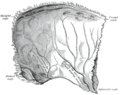
 Superior sagittal sinus laid open after removal of the skull cap. The chordæ Willisii are clearly seen. The venous lacunæ are also well shown; from two of them probes are passed into the superior sagittal sinus. | |
| Details | |
| Source | superior cerebral veins |
| Drains to | confluence of sinuses |
| Identifiers | |
| Latin | sinus sagittalis superior |
| MeSH | D054063 |
| TA98 | A12.3.05.109 |
| TA2 | 4856 |
| FMA | 50767 |
| Anatomical terminology | |
The superior sagittal sinus (also known as the superior longitudinal sinus), within the human head, is an unpaired area along the attached margin of falx cerebri. It allows blood to drain from the lateral aspects of anterior cerebral hemispheres to the confluence of sinuses. Cerebrospinal fluid drains through arachnoid granulations into the superior sagittal sinus and is returned to venous circulation.
Anatomy
Commencing at the foramen cecum, through which it receives a vein from the nasal cavity, it runs from anterior to posterior, grooving the inner surface of the frontal, the adjacent margins of the two parietal lobes, and the superior division of the cruciate eminence of the occipital lobe. Near the internal occipital protuberance, it drains into the confluence of sinuses and deviates to either side (usually the right). At this point it is continued as the corresponding transverse sinus.
It is triangular in section, narrow in front, and gradually increases in size as it passes backward.
Its inner surface presents the openings of the superior cerebral veins, which run, for the most part, obliquely forward, and open chiefly at the back part of the sinus, their orifices being concealed by fibrous folds; numerous fibrous bands (chordae Willisii) extend transversely across the inferior angle of the sinus; and, lastly, small openings communicate with irregularly shaped venous spaces (venous lacunae) in the dura mater near the sinus.
There are usually three lacunae on either side of the sinus: a small frontal, a large parietal, and an occipital, intermediate in size between the other two.
Most of the cerebral veins from the outer surface of the hemisphere open into these lacunæ, and numerous arachnoid granulations (Pacchionian bodies) project into them from below.
The superior sagittal sinus receives the superior cerebral veins, veins from the diploë and dura mater, and, near the posterior extremity of the sagittal suture, veins from the pericranium, which pass through the parietal foramina.
Clinical Significance
Cerebral Venous Oxygenation
Currently, the only available method for cerebral venous blood oxygenation monitoring is invasive and requires catheterization of the internal jugular vein. Researchers at University of Texas Medical Branch (UTMB) in Galveston, Texas are developing a noninvasive patient monitoring system that taps into the superior sagittal sinus to measure cerebral venous oxygenation[1].
Pulsing frequencies of near-infrared light are sent into the superior sagittal sinus vein. Hemoglobin in the blood absorbs the light at different frequencies depending on whether or not it is carrying oxygen. Absorption causes rapid thermal expansion of the hemoglobin resulting in a measurable acoustic wave. The software analyzes the signal and returns an absolute measurement of oxygen saturation.[2]
Additional images
-
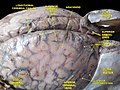
 Brain with sagittal sinus at centre, with various lacunae.
 -
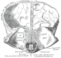
 Left parietal bone. Inner surface.
 -
 Frontal bone. Inner surface.
 -
 Base of the skull. Upper surface.
 -
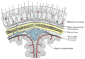
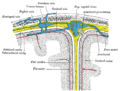 Diagrammatic representation of a section across the top of the skull, showing the membranes of the brain, etc.
 -
 Diagrammatic section of scalp.
 -
 Human brain dura mater
 -
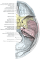
 Meninges and superficial cerebral veins.Deep dissection.Superior view.
 -
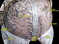 Meninges and superficial cerebral veins.Deep dissection.Superior view.


See also
External links
References
 This article incorporates text in the public domain from page 654 of the 20th edition of Gray's Anatomy (1918)
- ^ Petrov, I. Y.; Petrov, Y.; Prough, D. S.; Deyo, D. J.; Cicenaite, I.; Esenaliev, R. O. (2011-12-14). "Optoacoustic monitoring of cerebral venous blood oxygenation through extracerebral blood". Biomedical Optics Express. 3 (1): 125–136. doi:10.1364/BOE.3.000125. ISSN 2156-7085. PMC 3255330. PMID 22254173.
- ^ Petrova, I. Y.; Petrov, Y. Y.; Esenaliev, R. O.; Deyo, D. J.; Cicenaite, I.; Prough, D. S. (2009-04-27). "Noninvasive monitoring of cerebral blood oxygenation in ovine superior sagittal sinus with novel multi-wavelength optoacoustic system". www.osapublishing.org. doi:10.1364/OE.17.007285. Retrieved 2015-10-29.