Azygos continuity

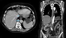
The azygos continuity of the inferior vena cava is one of the congenital malfunctions of the venous outflow from the lower half of the body. The inferior vena cava is not present throughout; the segment between the liver and kidneys is missing. As in normal anatomy, the hepatic veins drain almost directly into the right atrium via a short hepatic segment. However, the renal veins flow with the infrarenal segment of the inferior vena cava into the azygos vein , which is much wider than normal due to the increased blood flow. This then flows normally into the superior vena cava . There are a number of variations , each with differently designed vessel sections.
history
Descriptions of this anatomical peculiarity in combination with other malformations can be found by John Abernethy as early as 1793.
Emergence
It is believed that the absence of the anastomosis between the right subcardinal vein and hepatic veins leads to atrophy of the subcardinal vein.
frequency
In patients with no symptoms, the anomaly is occasionally discovered as an incidental finding in cross- sectional examinations. The prevalence is given as 0.6%. Azygos continuity occurs more frequently in the context of a heterotaxic syndrome .
Clinical significance
Whether symptoms are also associated with the misalignment and therefore a disease value depends on whether the existing bypass circuits are sufficient to ensure the return flow to the heart. Otherwise, thromboses and other consequences of chronic backflow such as B. Observe varices . If a syndrome occurs , other malformations, e.g. B. the heart are in the foreground.
Web links
Individual evidence
- ↑ a b c d Thomas Cissarek, Knut Kröger, Frans Santosa, Thomas Zeller (eds.): Vascular medicine therapy and practice . ABW Wissenschaftsverlag, 2009, p. 412.
- ^ J. Abernethy: Account of two instances of uncommon formation in the viscera of the human body . In: Philos Trans R Soc. , 83, 1793, pp. 59-66, jstor.org (PDF).
- ↑ a b J. E. Bass, MD Redwine, LA Kramer, PT Huynh, JH Harris: Spectrum of congenital anomalies of the inferior vena cava: cross-sectional imaging findings . In: Radiographics: a review publication of the Radiological Society of North America, Inc. Volume 20, Number 3, 2000 May-Jun, pp. 639-652, ISSN 0271-5333 . PMID 10835118 . (Review).
- ↑ S. Ginaldi, VP Chuang, S. Wallace: Absence of hepatic segment of the inferior vena cava with azygous continuation. In: Journal of computer assisted tomography. Volume 4, Number 1, February 1980, pp. 112-114, ISSN 0363-8715 . PMID 7354162 .
- ^ S. Müther, I. Dähnert: Das Heterotaxiesyndrom. In: Journal of Cardiac, Thoracic and Vascular Surgery. Volume 14, Number 3, June 2000, pp. 134-136, doi: 10.1007 / s003980070034
- ↑ Michalis Georgiadis: The "look behind the heart in the four-chamber view" and its importance for the sonographic diagnosis of fetal heart defects . (PDF; 846 kB). Inaugural dissertation. Rheinische Friedrich-Wilhelms-Universität, Bonn 2008.
