Dermatofluoroscopy
The Dermatofluoroskopie is a method to support the (early) diagnosis of skin cancer (malignant melanoma). The melanin in the skin tissue of suspicious skin areas (lesions) is examined and important information is provided from the upper layers of the skin. The underlying method uses an approach that enables a specific stimulation of the melanin fluorescence in the skin. This extremely weak fluorescence can be measured for the first time because it is no longer outshone by the fluorescence of the other, intensive endogenous fluorophores of the skin. The melanin fluorescence from melanocytes , nevus cells and melanoma cells shows characteristic spectral differences. The resulting fluorescence is visible and not outshone by the autofluorescence of the other, endogenous fluorophores in the cells. The transformation from benign nevus to malignant melanoma shows up as a characteristic redshift in the fluorescence spectrum. This spectral shift is the “universal fingerprint” of both nevus-based melanomas and de novo melanomas. The objective method differs fundamentally from other melanoma diagnostic methods, which are based, for example, on optically supported image recognition or the recognition of a changed morphology of the suspicious skin tissue. The process was patented in 2008.
Melanin fluorescence
The melanoma starts from the pigment-forming cells of the skin or mucous membrane, the so-called melanocytes. This is where melanin is formed, the complex pigment that is responsible for the brown color of the skin and UV protection. When energy is supplied to dyes, they go from the ground state to an excited state. They stay here for a short time before they return to their basic state. The dyes emit a characteristic fluorescence. In contrast to other molecules, melanin almost completely converts the added energy into heat; only about a ten-thousandth of the energy is available for fluorescence. Under conventional measurement conditions, this melanin fluorescence is completely outshone by the so-called autofluorescence of the skin, which emanates from its much more strongly fluorescent organic substances (e.g. NADH , flavin). If the fluorescence of melanin can be measured, it can be determined which cells are involved ( melanocytes , nevus cells or melanoma cells) without having to cut out the suspicious skin area.
Gradual 2-photon excitation
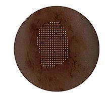
With the help of the gradual two-photon absorption it is possible to selectively stimulate melanin and make its fluorescence visible. For this purpose, nanosecond laser pulses with a wavelength of 800 nm are focused in the epidermis and the resulting melanin fluorescence is detected in the range from approx. 430 to 680 nm. Using this excitation technique, the lesion is scanned point by point with a step size of 200 µm. Degeneracies can be visualized in dermatofluoroscopy as a red shift in the spectrum of melanin fluorescence. An automated data analysis evaluates the extent of the spectral shift, the frequency and the spatial distribution and determines a score that either suggests removal (excision) of the examined lesion or a follow-up check or classifies the lesion as unsuspicious. Dermatofluoroscopy can be used not only on the patient but also on the histological specimen.
Approval and medical use
The method of dermatofluoroscopy is used in the dermatofluoroscope DermaFC . The class 2a medical device has been CE-certified since 2017 and approved for use as diagnostic support by dermatologists. The clinical evaluation of the DermaFC was carried out as part of a three-year, multi-center clinical trial at the University Clinic Tübingen , the University Clinic Heidelberg and the Charité Berlin. This exam was successfully completed in July 2017.
Individual evidence
- ^ A b Matthias Scholz, Dieter Leupold: Patent: LTB Lasertechnik Berlin GmbH. 2008. Spatially resolved measuring method for the detection of melanin in fluorophore mixtures in a solid sample . DE. 10 2006 029 809.8. February 8, 2008.
- ↑ J. Welzel, E. Sattler: New melanoma diagnostics on the basis of selectively excited melanin fluorescence by means of stepwise two-photon excitation . In: Non-invasive physical diagnostics in dermatology . Springer Verlag, 2016, pp. 119–122.
- ↑ Dieter Leupold, Matthias Scholz, G. Stankovic et al .: "The stepwise two-photon excited melanin fluorescence is a unique diagnostic tool for the detection of malignant transformation in melanocytes." Pigment Cell Melanoma Res . 2011 Jun; 24 (3): 438-45.
- ↑ Sarah Freudenberger: Information content of the melanin-dominated fluorescence of cutaneous melanocytic tumors in paraffin: Statements in relation to the histological findings . Ed .: Tübingen, Univ. 4 Faculty of Medicine [10874], Tübingen 2014.
