diastole
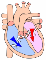
The diastole of the chambers of the heart ( Greek διαστολή "the expansion") is the relaxation and filling phase , in contrast to the systole , the tension and expulsion phase. The diastole of the atria occurs during the systole of the ventricles.
Classification
Mechanically , the diastole begins with the relaxation of the ventricular muscles and simultaneous closure of the pocket valves to the large arteries. It ends with the end of the sail flaps and the reopening of the pocket flaps. In the ECG , this is the phase between the end of the T wave and the beginning of the Q wave. The phase between the beginning of the T wave and the beginning of the following P wave is sometimes referred to as the so-called “electrical diastole” . In other literature the electrical diastole is equated with the mechanical. Echocardiographically , the diastolic filling of the ventricles is characterized by E and A waves.
Mechanical diastole is divided into four phases:
-
Relaxation phase (isovolumetric relaxation): period after the contraction of the heart chambers in which both the pocket and cusps are closed
- ECG: End of the T wave to the middle of the TP segment
- Echo: End of systolic outflow to start of E-wave
-
Early filling phase ( "active diastole" ): The heart chambers suck in blood by stepping up the valve level via the open leaflets.
- ECG: middle of the TP segment to the beginning of the P wave
- Echo: E-wave
-
Diastasis , is sometimes still counted as part of the early filling phase.
- ECG: P wave
- Echo: phase between E-wave and A-wave
-
Late filling phase: contraction of the atria until they relax and the leaflet valves close with further filling of the ventricles
- EKG: PR route
- Echo: A wave
Disorders of diastole can be seen as a cardiac arrhythmia , e.g. B. atrial fibrillation , or as a restriction in the quality of the diastolic filling of the heart chambers. Initially without severe symptoms, but as the disorder increases, this can lead to reduced performance in everyday life due to shortness of breath during physical exertion and even heart failure, diastolic heart failure .
See also
literature
- Roche Lexicon Medicine . 5th edition. Elsevier, Urban & Fischer, Munich 2003, ISBN 3-437-15072-3 ( diastole , active diastole [accessed March 27, 2009]).
- I. Krakau, H.Lapp (Hrsg.): The heart catheter book - Diagnostic and interventional catheter technology . 2nd Edition. Thieme, Stuttgart / New York 2005, ISBN 3-13-112412-1 , p. 95-96 ( books.google.de ).
Individual evidence
- ↑ C. Fahlke et al .: Pocket Atlas Physiology . 1st edition. 2008, ISBN 978-3-437-41917-1 , p. 198
- ↑ electrical diastole . In: Roche Lexicon Medicine . 5th edition. Elsevier, Urban & Fischer, Munich 2003, ISBN 3-437-15072-3 ( Gesundheit.de [accessed March 27, 2009]).
- ↑ a b v. Olshausen: EKG information . 8th edition. Steinkopf, Darmstadt 2005, ISBN 3-7985-1408-9 , pp. 2–3.
- ↑ Mewis, Riessen, Spyridopoulos (ed.): Cardiology compact - Everything for ward and specialist examination . 2nd Edition. Thieme, Stuttgart / New York 2006, ISBN 978-3-13-130742-2 , pp. 232 + 234 ( books.google.de ).
- ^ Flachskamp: Kursbuch Echokardiographie . 2nd Edition. Thieme, Stuttgart / New York 2000, ISBN 3-13-125672-9 , pp. 128 ( limited preview in the Google book search - the 3rd edition from 2006 is linked).

