Heart valve
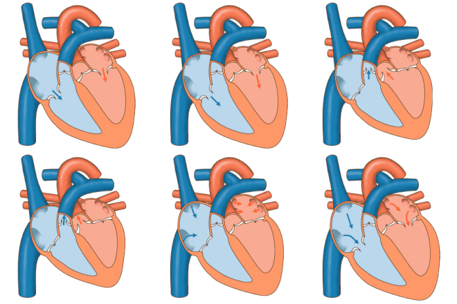
The four heart valves (Latin: valva = flap; valvula = small flap) act as valves in the heart and prevent the blood from flowing back in the wrong direction.
Overview
Each half of the heart has a leaflet valve ( atrioventricular valve ) and a pocket valve ( semilunar valve ). The leaflet valves lie between the atrium and ventricle and are called the bicuspid valve or mitral valve (left) and tricuspid valve (right). The pocket valves are located between the chamber and the outflow vessel and are called the pulmonary valve (right) and aortic valve (left).
The aortic valve forms together with the pulmonary valve in the 5th to 7th week of embryonic development . Heart valves are made up of folds of the endocardium that open and close concentrically. Normal heart valves do not represent any significant resistance to the blood flow, as this simply presses them against the heart wall when open. The return flow that then sets in unfolds the heart valves and thus closes the flow path. On the leaflet valves, the valve leaflets are prevented from turning in the wrong direction by tendon threads , which are tightened by the papillary muscles of the respective heart chamber during systole .
The pocket flaps cause the second heartbeat when they strike .
The flaps and their parts
Tricuspid valve
The "inlet valve" between the right atrium and the right ventricle is called the tricuspid valve ( valva atrioventricularis dextra ). It usually consists of three ( tri ) sails ( cuspis = tip, point ):
- Cuspis angularis (cranio-medial)
- Cuspis parietalis (lateral)
- Cuspis septalis (caudal).
Pulmonary valve
The "outlet valve" between the right ventricle and the pulmonary circulation is called the pulmonary valve ( Valva trunci pulmonalis ). It is designed as a pocket flap with normally three crescent-shaped flap pockets ( valvulae semilunares ) that prevent the backflow of blood into the right ventricle during diastole. During systole, the valve opens as soon as the pressure in the right ventricle exceeds that in the pulmonary artery.
Mitral valve (bicuspid valve)
The "inlet valve" between the left atrium ( atrium sinistrum ) and the left ventricle ( ventriculus sinister ) is called the bicuspid valve ( valva atrioventricularis sinistra ). This valve has two ( bi ) valve leaflets ( cuspides , singular cuspis ):
- Cuspis anterior (in veterinary anatomy: cuspis septalis )
- Cuspis posterior (in veterinary anatomy: cuspis parietalis )
Its shape is reminiscent of a miter and is therefore also known as the mitral valve ( valva mitralis ).
Aortic valve

Aortic Valve = Aortic Valve
Bicuspid Valve = Mitral
Valve Pulmonary Valve = Pulmonary Valve
Tricuspid Valve = Tricuspid Valve
The "outlet valve" of the left ventricle to the aorta is the aortic valve ( valva aortae ). It is designed as a pocket flap with three pockets ( valvulae semilunares ).
- Valvula semilunaris dextra or right pocket
- Valvula semilunaris sinistra or left pocket
- Posterior semilunar valvula or posterior pocket
People with a congenital two-pocket aortic valve are at increased risk of aortic valve stenosis and aortic dissection .
Heart valve diseases
- Main article: Valvular heart disease
Constrictions of the heart valves are used as stenoses (→ congenital aortic valve stenosis and acquired aortic valve stenosis , Trikuspidalklappenstenose , pulmonary valve stenosis , mitral valve stenosis ) and final inabilities as insufficiencies (→ tricuspid valve insufficiency , pulmonary valve , aortic regurgitation , mitral regurgitation hereinafter). Valve defects are the most common cause of abnormal heart murmurs . Under certain circumstances, they can be repaired with a heart valve reconstruction.
When inflammation of the endocardium can to shrink, scarring and "bonding" of one or more flaps come, can result in both valvular and valvular insufficiency.
If a heart valve is deformed from birth or has changed pathologically in the course of life, it can be replaced by an artificial heart valve (see heart valve replacement ).
literature
- Uwe Gille: Cardiovascular and immune system, Angiologia. In: Franz-Viktor Salomon, Hans Geyer, Uwe Gille (Ed.): Anatomy for veterinary medicine. 2nd, revised and expanded edition. Enke, Stuttgart 2008, ISBN 978-3-8304-1075-1 , pp. 404-463.


