Heart sound
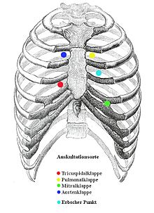
As heart sounds are called during the heart action arising audible vibrations (15-400 Hz) that the chest wall be transferred. With the ear or the stethoscope on , two of the four heart sounds can be heard ( auscultation , e.g. at Erb's point ).
- The 1st heart sound is dull and lasts 0.14 s. It comes about because the ventricular muscles contract around the incompressible blood when the leaflet valves ( heart valves between the atrium and ventricle) close ("muscle tension tone"). It can be heard best above the apex of the heart. The earlier definition of the 1st heart sound as "closure of the atrioventricular valves" could not be maintained physiologically. In the normal heart, the closing or opening of the atrioventricular valves themselves cannot be heard.
- The 2nd heart sound is lighter, louder and shorter (0.11 s) than the 1st heart sound. It is caused by the vibration of the blood column in the vessels immediately after the closure of the pocket valves of the aorta and pulmonary trunk ("valve closing tone"). It is best heard above the heart base.
In order to distinguish the first from the second heart sound in absolutely arrhythmic patients, the pulse can be felt at the same time during auscultation (for example the wrist pulse on the radial artery ). The first heart sound immediately precedes the perceived pulse wave.
Physiological (normal) heart sounds
In phonocardiogram four heart sounds can be differentiated:
- I. Heart tone in the tension phase of the heart (R-wave in the ECG )
- II. Heart sound when the pocket flaps of the two arteries of the heart are closed (shortly after the T wave in the ECG)
- III. Heart tone in the early filling phase due to the blood flowing into the heart chamber (falls in the front third of the TP segment in the ECG)
- IV. Heart sound that is only occasionally found as the sound of the atrial contraction (shortly after the P-wave on the EKG).
The 3rd and 4th heart sounds are only physiological to a certain extent in children and adolescents; in older patients they are always to be regarded as pathological. The 3rd heart sound comes from the impact of the blood stream on the stiff wall of the (insufficient) heart chamber; the presence of a third heart sound (and the resulting “protodiastolic gallop rhythm”) is of prognostic significance in heart failure and acute myocardial infarction . The 4th heart tone (also called atrial tone) is caused by the increased atrial contraction when the ventricle is more difficult to fill. If the 3rd and 4th heart sound are present at the same time, a "summation gallop" can occur in the event of a rapid heartbeat (tachycardia).
The mitral opening tone (MOED) may be heard in the early diastole 0.08–0.1 s after the 2nd heart tone in patients with narrowing of the mitral valve ( mitral stenosis ). This is usually followed by the typical "low-frequency diastolic rumble". A tricuspid opening sound is caused by increased filling pressure on the AV valves of the right heart, for example in the case of tricuspid stenosis or atrial septal defect. In addition, AV valve opening tones can occur when the circulating volume and the speed of early diastolic blood flow into the heart chambers are increased.
Pathological (abnormal) heart murmurs
If heart murmurs are added to the heart tones , these, in contrast to the heart tones, usually indicate heart valve defects . For example, a heart murmur during systole - the tension phase and outflow phase of the heart - is mostly due to a stenosis (narrowing) of the pocket valves or an insufficiency (leakage) of the leaflet valves. A diastolic noise - during the relaxation phase and the filling phase of the heart - usually indicates leaky pocket valves or narrowed leaflet valves. A stethoscope is used to listen to normal and pathological heart sounds and murmurs. It should be noted that the pathological heart sounds, i.e. 3rd and 4th heart sounds, are low-frequency and can therefore be heard much better with the so-called funnel of the stethoscope than with the membrane.
See also
literature
- Klaus Holldack, Klaus Gahl: Auscultation and percussion. Inspection and palpation. Thieme, Stuttgart 1955; 10th, revised edition, ibid. 1986, ISBN 3-13-352410-0 , pp. 104–128.
Web links
- Auscultation of the lungs and heart
- Heart murmurs and heart sounds to watch and listen to. Heilpraktikerausbildung24.de
