Fluorescence spectroscopy
The fluorescence spectroscopy is a spectroscopic technique of analytical chemistry . It uses fluorescence phenomena for the qualitative and quantitative analysis of substances.
Physical basics
As fluorescence is called the light-emission by prior absorption of a photon, if the decay time of the radiation is very short (i. E., The lifetime of the excited state is of the order 1-100 nanoseconds ). According to the following equation, the intensity of the fluorescence radiation is directly proportional to the intensity of the excitation radiation.
With
- : "Intensity" of the fluorescence radiation (emitted photons per time and area)
- : Fluorescence quantum yield (= number of emitted photons / number of absorbed photons)
- : "Intensity" of the excitation radiation (irradiated photons per time and area)
- : molar decadic extinction coefficient
- : Concentration
- : Layer thickness of the cuvette
This formula is only valid for weak absorption, i.e. H. , so that
(1) only a negligible fraction of the emitted photons are absorbed again
(2) for the fraction of the absorbed photons the following applies:
The excitation needs more energy, so the fluorescence spectra are shifted to longer wavelengths ( Stokes shift ). Molecules that have vibration levels that are suitably spaced apart can, if they are sufficiently close, take over the energy without radiation and thus lead to fluorescence quenching. A standard addition can be used to take these effects into account .
Device structure
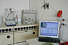
The structure of a fluorimeter is similar to that of a photometer , but the fluorescence with a certain emission wavelength ( ) is always measured at an angle of 90 ° so as not to include the excitation radiation ( ). In contrast to photometry, the radiation intensity of the emission is measured perpendicular to the direction of the excitation rays. An emission monochromator is connected upstream of the fluorimeter in order to remove residues of the excitation light ( scattered light ). High-pressure gas discharge lamps are usually used as the light source. Laser beams have also been used increasingly since the 1970s.
See also
literature
- Schwedt: Analytical Chemistry . 2nd Edition. WILEY-VCH, 2008, ISBN 978-3-527-31206-1 .
Web links
Individual evidence
- ↑ P. Atkins, Short textbook physical chemistry, 3rd edition, Wiley-VCH, 2002.
- ^ Gey: Instrumental and analytics and bioanalysis . 2nd Edition. Springer, 2008, ISBN 978-3-540-73803-9 , doi : 10.1007 / 978-3-540-73804-6 .


