Havers Canal
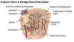
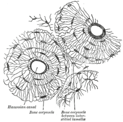
A Havers canal (Syn. Havers canal) is the central canal in the middle of an osteon , the basic functional unit in the bone cortex ( substantia compacta ) . It is concentrically enclosed by 5 to 20 µm thick bone lamellae. Inside are small blood vessels with fenestrated endothelium ( sinusoids ) and often non- myelinated nerve fibers. A Haversian canal thus serves to supply nutrients and transmit stimuli. The bone cells ( osteocytes ) are connected to the Haversian canal via so-called canaliculi . Adjacent Havers canals are connected to one another via Volkmann canals .
The British doctor Clopton Havers (1657–1702) discovered the canals named after him in 1691.
literature
- Clopton Havers: Osteologia nova, sive novae quedam observationes de ossibus et partibus ad illa pertinentibus ... Francofurti & Lipsiae, Apud Georgium Wilhelmum Kuhnium, 1692.
- Clopton Havers: A Short Discourse Concerning Concoction: Read at a Meeting of the Royal Society. In: Philosophical Transactions. 21/1699, pp. 233-47.
- S. Mohsin: Three dimensional reconstruction of haversian canals using X-ray micro-computed tomography. In: Bone. 38/2006, p. 16.
- I. Albu et al .: Haversian canals anastomoses in the long human bones. In: Morphologie et embryologie. 33/1987, pp. 167-9.
- C. Veleanu et al .: A simple and effective technique for haversian canals injection. In: Folia Morphologica . 21/1973, pp. 27-30.
- H.-J. Wagner: Basic features of molecular histology - cartilage and bone tissue. Anatomical Institute of the University of Tübingen

