Sinusoid (blood vessel)

A sinusoid is a small blood vessel that corresponds to an enlarged capillary with an average diameter of 10-30 µm and, due to its discontinuous endothelium, is completely permeable to all plasma components . For this reason, sinusoids are often referred to as discontinuous capillaries . Sinusoids occur, for example, in the liver as so-called liver sinusoids , in the spleen or in the bone marrow .
construction
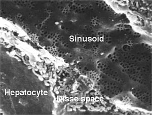
While blood vessels normally have a continuous endothelium with a continuous basal lamina , sinusoids are characterized by a missing or incomplete basal lamina. In the endothelium of the sinusoids of the liver and bone marrow, there are transcellular pores that are approximately 0.1 µm in diameter in the liver and up to 3 µm in diameter in the bone marrow. In the spleen, on the other hand, there are slits between the longitudinally oriented endothelial cells; the basement membrane is missing except for short sections that make contact with the pulp cords as part of the ring fibers.
function
The structure enables the exchange of larger molecules and sometimes cells between tissue and blood, for example the transfer of plasma proteins synthesized in the liver or the re-entry of erythrocytes from the open circulation in the spleen .
Individual evidence
- ^ LCU Junqueira, José Carneiro: Histology . Springer Verlag, 2005, 6th edition, ISBN 3-540219-65-X , page 275.
- ↑ Ulrich Welsch: Textbook Histology . Sobotta Verlag, 2006, 2nd edition, ISBN 978-3-437-44430-2 , pages 226, 246, 395.
- ^ Rolf Kötter: Wall of the liver sinusoids. Anatomie.net, accessed June 11, 2014 .
- ↑ a b L.CU Junqueira, José Carneiro: Histology . Springer Verlag, 2005, 6th edition, ISBN 3-540219-65-X , pages 181-185.