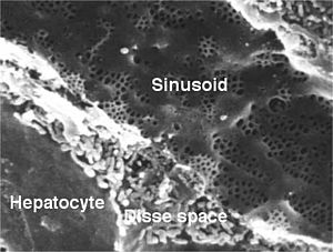
Liver sinusoid

Sinusoid from the liver of a rat with fenestrated endothelial cells . The diameter of the fenestrae is approx. 100 nm and that of the sinusoids approx. 5 µm . Scanning electron micrograph by Robin Fraser, University of Otago .
A liver sinusoid ( intralobular vein ) is an enlarged capillary vessel ( sinusoid ) that transports the oxygen-rich blood from the hepatic artery ( arteria hepatica propria ) and the nutrient-rich blood from the portal vein ( vena portae ). The sinusoids have Kupffer cells , which can absorb and destroy foreign substances (e.g. bacteria ) .
Individual evidence
- ↑ Ulrich Welsch: Textbook Histology . Sobotta Verlag, 2006, 2nd edition, ISBN 978-3-437-44430-2 , pages 226, 246, 395.
- ^ Rolf Kötter: Wall of the liver sinusoids. Anatomie.net, accessed June 11, 2014 .
- ^ LCU Junqueira, José Carneiro: Histology . Springer Verlag, 2005, 6th edition, ISBN 3-540219-65-X , pages 181-185.