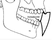
Posselt diagram
The Posselt diagram ( English Posselt's Envelope of Motion ) shows the limit movements of the incisal point of the lower jaw in the sagittal plane . The movement possibilities of the lower jaw can only take place within certain spatial limits, which are determined by the anatomy of the temporomandibular joints , the morphology of the rows of teeth , the viscoelastic properties of the muscles , the joint ligaments and the skin .
Limit positions of the Posselt diagram
From the patient's own occlusion position , the lower jaw can be actively moved forward. It slides with the incisal edges of the lower incisors on the palatal guide surfaces of the upper anterior teeth into the incisal edge position of the anterior teeth.
By further contraction of the masticatory muscles, the lower jaw can be moved into the greatest possible advance position, the maximum protrusion.
From the habitual intercuspation, the lower jaw can still be shifted slightly backwards in most people. The lower jaw slides into the retral contact position, which is also called the terminal hinge axis position.
From this limit position, the lower jaw can perform pure rotation or hinge movements up to a cutting edge distance of about 25 millimeters.
If the lower jaw is opened beyond the terminal hinge axis position, a sliding movement of the condyles begins by pulling the masticatory muscles directed forwards.
The opening movement ends at the position of the maximum mouth opening.
The border movements are for the
- Protrusion (advancing movement) 7–10 mm
- Opening 40-60 mm
- Laterotrusion (sideways movement) 10–15 mm
- Retrusion (backward thrust) 0-2.2 mm.
designation
The Posselt diagram is named after the Swedish dentist Ulf Posselt (1914–1966). He worked in the prosthetics department at Karolinska Institutet , Royal Dental College, Stockholm, in the anatomical institute at Lunds universitet and in the X-ray diagnostics department at the State Dental School, Malmö.
literature
- Ulf Posselt, Physiology of occlusion and rehabilitation, Blackwell Scientific, 1962
Individual evidence
- ↑ Bernd Reitemeier: Introduction to dentistry . Georg Thieme Verlag, 2006, ISBN 978-3-13-139191-9 , pp. 101-102.
- ^ Ulf Posselt: Movement areas of the mandible. In: The Journal of Prosthetic Dentistry. 7, 1957, p. 375, doi : 10.1016 / S0022-3913 (57) 80083-3 .
- ^ JA Salzmann: Ulf Posselt, Studies in the mobility of the human mandible. In: American Journal of Orthodontics. 39, 1953, p. 471, doi : 10.1016 / 0002-9416 (53) 90062-1 .
- ↑ Ulf Posselt, Odont. D .: Range of movement of the mandible. In: The Journal of the American Dental Association. 56, 1958, p. 10, doi : 10.14219 / jada.archive.1958.0017 .
- ↑ Wolfgang Gühring, Joachim Barth: Anatomie: special biology of the masticatory system . Verlag Neuer Merkur GmbH, 1992, ISBN 978-3-921280-84-3 , pp. 262-263.


