
Six year mol

The term six-year molar names the first large molar tooth ( molar , Latin molaris , millstone). The name comes from the fact that the first large molars in children erupt into the oral cavity around the age of six. They appear without a milk tooth falling out beforehand . In the front permanent teeth, one milk tooth falls out before they erupt. Not so with the large molars. They make their way as the first permanent teeth behind the last milk teeth, often unnoticed by children and parents. This process is therefore called incremental toothing . In the FDI tooth scheme , the six-year molars are designated 16, 26, 36, and 46.
Interlocking
The six-year molars have a central position in the human dentition. They have the largest chewing surface. With the emergence of the first toothing of the first molars, different bite positions can occur, which influence the toothing of the remaining teeth. In the tooth scheme , the six-year molars have the tooth designations 16, 26, 36, 46 . They are also called “sixes” (6s) because they represent the sixth tooth, calculated from the center. The six-year-old molars need several months of tooth eruption to reach the occlusal plane. Both the position of the molars and the poor opening of the child's mouth make it difficult to clean the teeth , which is why these important teeth have the highest levels of caries . This can be prevented by sealing the teeth immediately after the breakthrough.

Upper jaw bite. behind it the six-year molars break through

Dental scheme; Six-year molars (red), milk teeth on the right
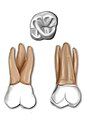
Maxilla six-year molar

Carabelli's tubercle

The tooth equator , the largest circumference of the six-year molar with infra and supra curvature


anatomy

upper jaw
The six-year molars in the upper jaw usually consist of four cusps, two on the buccal side (towards the cheek) and two on the palatal side (towards the palate). Every third six-year molar has a small fifth cusp on the mesio-palatal cusp, the tuberculum carabelli .
The upper first molars usually have three roots. The mesio-buccal root becomes wider buccolingually and has prominent depressions or ridges on its mesial and distal surfaces. The internal canal morphology is very variable, but the majority of the mesiobuccal roots contain two canals. The distobuccal root is generally rounded or oval in cross section and typically contains a single root canal. The palatal root is usually wider distally than bucco-lingual and ovoid in shape and usually contains only a single root canal. Although the palatal root generally appears straight on radiographs, it usually has a hump curvature in the apical third. Depressions on the cheek and palatal surfaces of the palate may be present, but are usually flat. The average total length of the upper first molar is 20.5 mm with a mean crown length of 7.5 mm and an average root length of 13 mm.
Lower jaw
The lower first molars have five cusps. From the occlusal view, the lower first molars have a pentagonal shape that tapers lingually. Both roots taper apically but are more prominent at the mesial root. The mesial root is wider buccolingually and its tip is blunt. The largest circumference is located buccally at the upper end of the apical third of the tooth crown, with the course of the occlusal two-thirds of the surface being flat. Lingually, the greatest circumference is in the middle third of the tooth. The lingual surface is uniformly convex.
pathology
The lower first molars are the most common, the upper first molars the second most common carious teeth, and those that most often undergo endodontic treatment or extraction . Up to 21% of all extracted teeth are upper first molars. In the Hutchinson triad , the six-year-old molars are affected in 22–65% of cases, which are called Hutchinson teeth and are mulberry-shaped. The molar-incisor-hypomineralisation (MIH) is a special form of melt formation disorder, namely a systemically related hypomineralisation the first permanent molars and / or of the upper permanent incisors ( incisors ). It is a variant of the structural disorders of the hard tooth substance ( tooth enamel ).
Individual evidence
- ↑ Bernd Reitemeier: Introduction to dentistry . Georg Thieme, 2006, ISBN 978-3-13-139191-9 , p. 87 ( google.com ).
- ^ A b J. Craig Baumgärtner, John I. Ingle, 2008, Ingle's Endodontics. Hamilton, Ontario. BC Decker Inc. ISBN 0-19-920409-8 , ISBN 1-55009-333-9 .
- ^ Y. Zadik, V. Sandler u. a .: Analysis of factors related to extraction of endodontically treated teeth. In: Oral surgery, oral medicine, oral pathology, oral radiology, and endodontics. Volume 106, number 5, November 2008, pp. E31-e35, doi: 10.1016 / j.tripleo.2008.06.017 , PMID 18718782 .
- ↑ O. Braun-Falco, Gerd Plewig, HH Wolff: Dermatology and Venereology . Springer, 2013, ISBN 978-3-662-00524-8 , pp. 91 ( google.com ).
- ^ F. Crombie, D. Manton, N. Kilpatrick: Aetiology of molar-incisor hypomineralization: a critical review. In: International journal of pediatric dentistry / the British Pedodontic Society [and] the International Association of Dentistry for Children. Volume 19, Number 2, March 2009, pp. 73-83, doi: 10.1111 / j.1365-263X.2008.00966.x , PMID 19250392 . (Review).
- ↑ S. Laisi, H. Kiviranta et al. a .: Molar-incisor-hypomineralization and dioxins: new findings. In: European archives of pediatric dentistry: official journal of the European Academy of Pediatric Dentistry. Volume 9, Number 4, December 2008, pp. 224-227, PMID 19054476 .