Cervical sinus
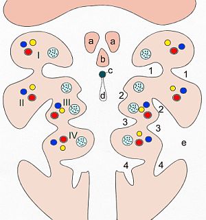
The cervical sinus is an invagination lined with ectodermal tissue on the outside of the cervical region (branchial region, "gill intestine") of mammalian embryos. The second, third and fourth throat furrows open into the cervical sinus. It arises (in humans in the fourth week of pregnancy) when the mesenchyme of the second pharyngeal arch expands through increased growth kaudad until it merges with the anlage of the heart. This outgrowth is called the operculum (Latin: "lid") because it overlaps the cervical sinus like a lid. After the operculum has fused with the heart bulge, the cervical sinus is now tied off from the outer surface and is then referred to as the cervical vesicle . Sinus cervicalis or vesicula cervicalis are so-called transitory structures, which means that they regress in the course of further embryonic development and normally disappear completely.
Clinic in humans
If there are developmental defects in connection with the cervical sinus, so-called lateral branchiogenic neck cysts or neck fistulas develop on the anterior (ventral) edge of the sternocleidomastoid muscle . The cause of the formation of cervical cysts is incomplete regression of the cervical vesicle. If the operculum does not completely fuse with the heart bulge, this leads to throat fistulas. A distinction is made between the more frequent external and the rarer internal branchiogenic fistulas.
literature
- Ulrich Drews: Pocket Atlas of Embryology. Georg Thieme Verlag, Stuttgart 2006, ISBN 3-13-109902-X .
- Compact anatomy textbook. Volume 3: Internal Organ Systems. Schattauer Verlag, Stuttgart 2004, ISBN 3-7945-2063-7 .
- P. Kaufmann, H. Leisten, U. Mangold: The development of the gill arch in the rat and mouse. In: Acta Anatomica . Volume 110, 1981, pp. 7-22, doi: 10.1159 / 000145408 .
