Synergist (muscle)
Synergist ( Greek συνεργεῖν synergeín , 'to work together') is a muscle called in medicine that supports, strengthens or enables the movement of another muscle. This definition cannot be applied to a muscle in general, but always refers exclusively to a specific movement. So operating in different movements of the pectoralis major , for example, times of triceps brachii muscle (z. B. bench press or push ups) synergistically, but other of biceps brachii (z. B. Butterfly).
A distinction is made between “direct” and “indirect” synergists.
Direct synergists aid the agonist's work by performing the same or a very similar movement. For example, three muscles are involved in flexion in the elbow joint , the biceps brachii , brachial and brachioradialis muscles, which have different effects depending on the position of the joint, but are almost always direct synergists of the others.
Indirect synergists make the work of the agonist possible in that they fix bone points at which the agonist inserts. So z. For example, lifting the leg while walking is impossible without the indirect synergistic effect of the abdominal muscles (especially the rectus abdominis muscle ) that fix the pelvis . Without the fixation of the pelvis, contraction of the leg lifters would cause the pelvis to tilt forward and not raise the leg. These muscle reactions are controlled by postural reactions of the brain stem , which combine “static and dynamic, highly complex sensorimotor basic building blocks”. The more precisely these postural synergies work together, the smoother, safer and more effortless the movement appears. The interaction of the synergists can be trained and refined through many repetitions.
The knowledge of muscular synergists (especially the indirect synergists) is crucial for all people who work actively or passively with movement, e.g. B. athletes, physiotherapists, trainers, sports doctors, etc., since the failure of a synergist to perform certain (frequently recurring) movements can often be the reason for the occurrence or maintenance of complaints.
Example external eye muscles
Each eye has six outer eye muscles that are responsible for its coordinated movements. Two of them each have a similar muscle level and turn the eye around an almost identical axis of rotation, but each in an opposite direction of rotation. These muscles are called antagonists . In contrast, muscles that move the eye around a similar axis of rotation in the same direction are called synergists. This terminology is also used when only partial functions of the respective muscles agree or counteract one another. It is only fully applicable for ductions, i.e. movements of one eye. If one extends the consideration to the opposite eye by the description of contralateral synergists and antagonists in the execution of binocular eye movements, then this definition for vergences, opposing eye movements, must be restricted.
- Ipsilateral (equilateral) synergists and antagonists with regard to the respective muscle function
| Agonist | function | Synergists | Antagonists |
|---|---|---|---|
| M. rect. Medialis | Adduction |
Rect. Superior, rect. Inferior |
M. rect.lateralis, M. obl. Superior, M. obl. Inferior |
| Lateral rectus muscle | Abduction |
M. obl. Superior, M. obl. Inferior |
M. rect.medialis, M. rect. Superior, M. rect. Inferior |
| M. rect. Superior | Elevation
Inside roll Adduction |
M. obl. Inferior
M. obl. Superior M. rect.medialis, M. rect.inferior |
M. rect.inferior, M. obl. Superior
M. rect. Inferior, M. obl. Inferior M. rect.lateralis, M. obl. Superior, M. obl. Inferior |
| M. rect. Inferior | Lowering
Outside roll Adduction |
M. obl. Superior
M. obl. Inferior Medial rectus muscle, superior rectal muscle |
M. rect. Superior, M. obl. Inferior
M. obl. Superior, M. rect. Superior M. rect.lateralis, M. obl. Superior, M. obl. Inferior |
| M. obl. Superior | Lowering
Inside roll Abduction |
M. rect. Inferior
M. rect. Superior M. rect.lateralis, M. obl. Inferior |
M. rect. Superior, M. obl. Inferior
M. rect. Inferior, M. obl. Inferior M. rect.medialis, M. rect. Superior, M. rect. Inferior |
| M. obl. Inferior | Elevation
Outside roll Abduction |
M. rect. Superior
M. rect. Inferior M. rect.lateralis, M. obl. Superior |
M. rect.inferior, M. obl. Superior
M. obl. Superior, M. rect. Superior M. rect.medialis, M. rect. Superior, M. rect. Inferior |
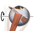
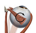
- Graphic representation of the involvement of individual muscles (synergists) in the respective rotational movements using the example of the right eye
| Elevation. The main participants are the M. rectus superior and the M. obliquus inferior | Lowering. The main participants are the M. rectus inferior and M. obliquus superior | Adduction. The main involved is the M. rectus medialis and the vertical Mm. recti | Abduction. The main involved is the lateral rectus muscle and the Mm. obliqui | Inside roll. The main participants are the M. obliquus superior and the M. rectus superior | Outside roll. The main participants are the M. obliquus inferior and the M. rectus inferior |
See also
literature
- Th. Axenfeld (conception), H. Pau (ed.): Textbook and atlas of ophthalmology . With the collaboration of R. Sachsenweger and others Gustav Fischer Verlag, Stuttgart 1980, ISBN 3-437-00255-4 .
- Albert J. Augustin: Ophthalmology . Springer Verlag, Berlin 2007, ISBN 978-3-540-30454-8 .
- Karl-Michael Haus: Neurophysiological treatment in adults: basics of neurology, treatment concepts, everyday therapy approaches. 2nd edition. Springer Verlag, 2010, ISBN 978-3-540-95969-4 .
Individual evidence
- ↑ Walther Grauman, Dieter Sasse: Compact textbook of the entire anatomy. Volume 4: Sensory systems, skin, CNS, peripheral pathways . 1st edition. Verlag Schattauer, 2004, ISBN 3-7945-2064-5 .
- ↑ Antje Hüter-Becker, among other things: biomechanics, kinetics, performance physiology, training theory. Georg Thieme Verlag, Stuttgart 2005, ISBN 3-13-136861-6 , p. 161.
- ↑ Herbert Kaufmann (with the collaboration of W. de Decker and others): Strabismus. Enke, Stuttgart 1986, ISBN 3-432-95391-7 , p. 38.


