Tooth movement
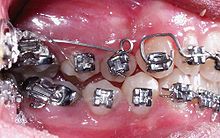
Tooth movements can be physiological, pathological and therapeutic. Physiologically, teeth are moved into their position by pressure on the cheeks and tongue. Pathologically, teeth can be moved by external influences (impact, impact) or occur in connection with periodontological diseases as tooth loosening . Tooth movement is used therapeutically as part of an orthodontic treatment to correct misaligned teeth ( English Orthodontic tooth movement ).
Basics of therapeutic tooth movement
Therapeutic tooth movement can occur due to the existing periodontal gap. Pressure application and elimination trigger special metabolic processes in the periodontal ligament. In principle, the bone is remodeled by osteoclasts and osteoblasts . This process is controlled by complex processes, which are the subject of numerous studies and research efforts. A variety of cytokines , proteins, and chemokines are discussed. For example, osteoclasts and pre-osteoclasts are regulated by different signal pathways and molecules. A key module for regulation is a protein called the receptor activator for the nuclear factor kB ligand ( RANK ligand ). To compensate for this, osteoprotegerin is secreted from the osteoblasts . The reaction to a certain force is very individual and depends on numerous factors, for example age, oral hygiene or smoking.
Phases of tooth movement
The tooth movement is divided into three phases:
- First phase: phase of initial damping. This lasts for about one to three days and the tooth is initially deflected and the blood circulation changes.
- Second phase: hyalinization . This phase lasts about 2–10 weeks. The tooth movement is reduced or comes to a complete standstill.
- Third phase: absorption . It is a direct bone resorption. The tooth movement is now accelerated.
Biological efficiencies
According to AM Schwarz, a distinction is made between four biological degrees of effectiveness.
- Subliminal forces: Do not lead to a change in tooth position
- Weak, short-term pressure forces: At 0.15-0.2 N / cm 2 , the blood flow in the capillaries is not blocked.
- Medium pressure forces: At 0.2–0.5 N / cm 2 , the capillary blood pressure is exceeded, but the tissue is not completely pressed.
- Fourth biological efficiency: With forces greater than 0.5 N / cm 2 , the periodontal area is pressed off, the consequences are often repairable, but in extreme cases the consequences can lead to tooth loss.
For a controlled tooth movement, therefore, forces of the second biological degree of effectiveness should be selected, if forces of the third degree of effectiveness are used, they should not act for longer than 8-12 hours.
Diagnosis
Tilted or elongated (seemingly elongated) teeth are characterized in the diagnostic chart with arrows that indicate the direction of kipping, elongation or tooth migration: → or ←, ↑ or ↓. A gap closure is indicated by two opposite brackets) (. If the gap has been closed by tooth migration , additional arrows are indicated before and / or after the brackets: →) (←.
In the following example, a gap closure is shown by tooth movement of the premolars 14, 24, 34 and 44 after an extraction that is necessary for orthodontics. A gap was closed between tooth 18 and 16 when tooth 17 was missing, in that tooth 18 had migrated in the mesial direction due to tooth migration.
| top right | top left | |||||||||||||||
|---|---|---|---|---|---|---|---|---|---|---|---|---|---|---|---|---|
| →) ( | ) ( | ) ( | Finding | |||||||||||||
| 18th | 17th | 16 | 15th | 14th | 13 | 12 | 11 | 21st | 22nd | 23 | 24 | 25th | 26th | 27 | 28 |
Tooth designation |
| 48 | 47 | 46 | 45 | 44 | 43 | 42 | 41 | 31 | 32 | 33 | 34 | 35 | 36 | 37 | 38 | |
| ) ( | ) ( | Finding | ||||||||||||||
| bottom right | bottom left | |||||||||||||||
Individual evidence
- ↑ OEMUS MEDIA AG: The control of orthodontic tooth movement . In: ZWP online - The news portal for the dental industry .
- ↑ Wichelhaus, Andrea: 2012 Color Atlases of Dentistry: Orthodontics - Therapy Volume 1 doi: 10.1055 / b-0034-18342
- ↑ Introduction to orthodontics - diagnostics, treatment planning, therapy. Bärbel Kahl-Nieke. 2009 Deutscher Ärzte-Verlag, ISBN 978-3-7691-3419-3 .
- ↑ Orthodontic dental technology (page 73). Edited by Friedbert Schmeil u. Ursula Hirschfelder. 2004. Verlag Neuer Merkur GmbH ISBN 3-929360-77-2 .
- ^ Rudolf W. Ott: Clinic and Practice Guide Dentistry . Georg Thieme Verlag, 2003, ISBN 978-3-13-131781-0 , p. 448.
