
Inferior epigastric vein: Difference between revisions
Content deleted Content added
Tom.Reding (talk | contribs) m +{{Authority control}} (1 source from Wikidata), WP:GenFixes on |
Fixed typo in ordinal Tags: Mobile edit Mobile web edit |
||
| (3 intermediate revisions by 3 users not shown) | |||
Line 1:
{{Use American English|date = January 2019}}
{{Short description|Large blood vessel}}
{{Use mdy dates|date = January 2019}}
{{Infobox vein
| Name = Inferior epigastric vein
Line 10 ⟶ 13:
| Artery = [[inferior epigastric artery]]
}}
In [[human anatomy]], '''inferior epigastric vein'''
==Additional images==
Line 22 ⟶ 25:
*[[Terms for anatomical location]]
*[[Hesselbach's triangle]]
== References ==
{{Reflist}}
==External links==
* {{SUNYAnatomyLabs|35|12|01|05}} - "Anterior [[Abdominal wall]]: Blood Vessels in the [[Rectus sheath]]"
* {{SUNYAnatomyFigs|35|04|07}} - "Incisions and the contents of the rectus sheath."
| |||
Revision as of 19:38, 11 April 2023
| Inferior epigastric vein | |
|---|---|
 Right inferior epigastric vein - view from inside of abdomen. | |
 The iliac veins. | |
| Details | |
| Drains from | superior epigastric vein |
| Drains to | external iliac vein |
| Artery | inferior epigastric artery |
| Identifiers | |
| Latin | vena epigastrica inferior |
| TA98 | A12.3.10.025 |
| TA2 | 5051 |
| FMA | 21162 |
| Anatomical terminology | |
In human anatomy, inferior epigastric vein are 1-2 veins accompanying the inferior epigastric artery. They drain into the external iliac vein just proximal to the inguinal ligament.[1]
Additional images
-
 The interfoveolar ligament, seen from in front.
 -
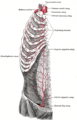 The internal mammary artery and its branches.
 -
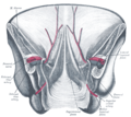 Posterior view of the anterior abdominal wall in its lower half. The peritoneum is in place, and the various cords are shining through.


See also
References
- ^ Standring, Susan (2020). Gray's Anatomy: The Anatomical Basis of Clinical Practice (42nd ed.). New York. p. 1251. ISBN 978-0-7020-7707-4. OCLC 1201341621.
{{cite book}}: CS1 maint: location missing publisher (link)
External links
- Anatomy photo:35:12-0105 at the SUNY Downstate Medical Center - "Anterior Abdominal wall: Blood Vessels in the Rectus sheath"
- Anatomy figure: 35:04-07 at Human Anatomy Online, SUNY Downstate Medical Center - "Incisions and the contents of the rectus sheath."
This cardiovascular system article is a stub. You can help Wikipedia by expanding it. |