
Superior mesenteric artery
| Superior mesenteric artery | |
|---|---|
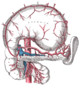
 The pancreas and duodenum from behind. (Superior mesenteric artery labeled at upper right.) | |
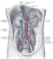
 Frontal view of the superior mesenteric artery and its branches. The large vessel (blue) beside the SMA is the superior mesenteric vein. A considerable number of different branching patterns exist. | |
| Details | |
| Source | abdominal aorta |
| Branches | inferior pancreaticoduodenal middle colic right colic intestinal branches (jejunal, ileal) ileocolic |
| Vein | superior mesenteric vein |
| Supplies | intestine |
| Identifiers | |
| Latin | arteria mesenterica superior |
| MeSH | D017538 |
| TA98 | A12.2.12.053 |
| TA2 | 4252 |
| FMA | 14749 |
| Anatomical terminology | |
In human anatomy, the superior mesenteric artery (SMA) arises from the anterior surface of the abdominal aorta, just inferior to the origin of the celiac trunk, and supplies the intestine from the lower part of the duodenum to the left colic flexure and the pancreas.
Location and path
It arises anterior to vertebra L1 in an adult. It is usually 1cm lower than the celiac trunk. It initially travels in an anterior/inferior direction, passing behind/under the neck of the pancreas and the splenic vein. Located under this portion of the superior mesenteric artery, between it and the aorta, are the following:
- left renal vein - travels between the left kidney and the inferior vena cava (can be compressed by the SMA at this location, leading to the so-called nutcracker syndrome.
- the third part of the duodenum, part of the small intestines
- uncinate process of the pancreas - this is a small part of the pancreas that hooks around the SMA
The SMA typically runs to the left of the similarly named vein, the superior mesenteric vein. After passing the neck of the pancreas it starts giving off its branches.
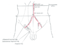
Branches
| Branch | Supplies |
| inferior pancreaticoduodenal artery | head of the pancreas and to the descending and inferior parts of the duodenum |
| middle colic artery | to the transverse colon |
| right colic artery | to ascending colon |
| intestinal arteries | branches to ileum, branches to jejunum |
| ileocolic artery | (terminal branch of the SMA) supplies last part of ileum, cecum, and appendix |
The middle, right, and ileocecal branches anastomose with each other to form a marginal artery along the inner border of the colon. This artery is completed by branches of the left colic which is a branch of the inferior mesenteric artery.
Pathology
- Compared to other vessels of similar size, the SMA is largely spared the effects of atherosclerosis. This is likely due to protective haemodynamic conditions
- Occlusion of the SMA almost invariably leads to intestinal ischemia and often has devastating consequences; up to 80% of SMA occlusions lead to death.[1]
- The SMA can compress the left renal vein, leading to the nutcracker syndrome and/or the third (horizontal) part of the duodenum, leading to SMA syndrome.
Additional images
-
 The celiac artery and its branches; the stomach has been raised and the peritoneum removed.
 -
 Abdominal part of digestive tube and its attachment to the primitive or common mesentery. Human embryo of six weeks.
 -
 Posterior abdominal wall, after removal of the peritoneum, showing kidneys, suprarenal capsules, and great vessels.
 -
 The anterior surfaces of the kidneys, showing the areas of contact of neighboring viscera.
 -
 Front of abdomen, showing surface markings for arteries and inguinal canal.


Reference
External link
- Anatomy figure: 39:02-01 at Human Anatomy Online, SUNY Downstate Medical Center - "Branches of the inferior mesenteric artery."
- Anatomy photo:40:11-0102 at the SUNY Downstate Medical Center - "Posterior Abdominal Wall: Branches of the Abdominal Aorta"
- Anatomy image:8008 at the SUNY Downstate Medical Center
- Anatomy image:8404 at the SUNY Downstate Medical Center
- Anatomy image:8815 at the SUNY Downstate Medical Center
- Anatomy image:8841 at the SUNY Downstate Medical Center
- Atlas image: abdo_wall70 at the University of Michigan Health System - "Posterior Abdominal Wall, Dissection, Anterior View"
- sup&infmesentericart at The Anatomy Lesson by Wesley Norman (Georgetown University)