
Rectus abdominis muscle
| Rectus abdominis | |
|---|---|
 The human rectus abdominis muscle. | |
| Details | |
| Origin | Pubis |
| Insertion | Costal cartilage of ribs 5-7, xiphoid process of sternum |
| Artery | inferior epigastric artery |
| Nerve | segmentally by thoraco-abdominal nerves (T7 to T12) |
| Actions | flexion of trunk/lumbar vertebrae |
| Antagonist | Erector spinae |
| Identifiers | |
| Latin | musculus rectus abdominis |
| MeSH | D017568 |
| TA98 | A04.5.01.001 A04.5.00.001 |
| TA2 | 2357 |
| FMA | 9628 |
| Anatomical terms of muscle | |
The rectus abdominis muscle is a paired muscle running vertically on each side of the anterior wall of the human abdomen (and in some other animals). There are two parallel muscles, separated by a band of connective tissue called the linea alba (white line). It extends from the pubic symphysis inferiorly to the xiphisternum and lower costal cartilages superiorly.
It is contained in the Rectus sheath.
The Rectus is crossed by fibrous bands, three in number, which are named the tendinous inscriptions. If well-defined, the rectus abdominis is colloquially called a "six-pack."
Function
[[Feline_Rectus_Abdominus.jpg|thumb|right|200px|A feline rectus abdominis muscle, from a common housecat. This specimen has some remaining fascia and also shows the external obliques.]] The rectus abdominis is a key postural muscle. It is responsible for flexing the lumbar spine, as when doing a 'sit-up'. The rectus abdominis can play a role in respiration in the event the patient is short of breath.
Blood supply
The inferior epigastric artery and vein (or veins) run superiorly on the posterior surface of the rectus abdominis, enter the rectus fascia at the arcuate line, and help to supply the muscle with blood.
Location
The Rectus abdominis is a long flat muscle, which extends along the whole length of the front of the abdomen, and is separated from its fellow of the opposite side by the linea alba.
It is much broader, but thinner, above than below, and arises by two tendons;
- the lateral or larger is attached to the crest of the pubis,
- the medial interlaces with its fellow of the opposite side, and is connected with the ligaments covering the front of the symphysis pubis.
The muscle is inserted by three portions of unequal size into the cartilages of the fifth, sixth, and seventh ribs.
The upper portion, attached principally to the cartilage of the fifth rib, usually has some fibers of insertion into the anterior extremity of the rib itself.
Some fibers are occasionally connected with the costoxiphoid ligaments, and the side of the xiphoid process.
Animals
The Rectus Abdominis is similar in most vertebrates. The most obvious difference between animal and human abdominal musculature is that in animals, there are a different number of tendinous intersections.
Additional images
-
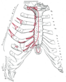 Anterior surface of sternum and costal cartilages.
 -
 The interfoveolar ligament, seen from in front.
 -
 Diagram of sheath of Rectus.
 -
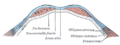 Diagram of a transverse section through the anterior abdomina wall, below the linea semicircularis.
 -
 The relations of the femoral and abdominal inguinal rings, seen from within the abdomen. Right side.
 -
 Horizontal disposition of the peritoneum in the lower part of the abdomen.
 -
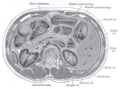 Transverse section through the middle of the first lumbar vertebra, showing the relations of the pancreas.
 -
 The left side of the thorax.
 -
 Surface anatomy of the front of the thorax and abdomen.
 -
 Muscles of the trunk
 -
This professional bodybuilder exhibits an easily visible six-pack.


External links
- Template:MuscleLoyola
- . GPnotebook https://www.gpnotebook.co.uk/simplepage.cfm?ID=168165454.
{{cite web}}: Missing or empty|title=(help) - Anatomy figure: 04:04-07 at Human Anatomy Online, SUNY Downstate Medical Center - "Muscles of the anterior chest wall with the pectoralis major muscles removed."
- Anatomy photo:18:01-0115 at the SUNY Downstate Medical Center - "Thoracic Wall: The Anterior Thoracic Wall"
- Anatomy figure: 35:06-07 at Human Anatomy Online, SUNY Downstate Medical Center - "Incision and reflection of the external abdominal oblique muscle."
- Anatomy figure: 35:07-01 at Human Anatomy Online, SUNY Downstate Medical Center - "Incision and reflection of the internal abdominal oblique muscle."
- Anatomy photo:35:10-0100 at the SUNY Downstate Medical Center - "Anterior Abdominal Wall: The Rectus Abdominis Muscle"
- Cross section image: pembody/body12a—Plastination Laboratory at the Medical University of Vienna
- Template:EMedicineDictionary
- Template:RocheLexicon
- Radiography at kumc.edu