Pubic bone

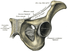
The pubic bone ( Latin os pubis ) is a flat, angular bone and part of the pelvis . It is present on both sides, with both pubic bones being connected in the midline by the pubic symphysis ( symphysis pubica ) - a fiber cartilaginous connection - so that the pelvic bones can move slightly towards each other. Upward and forward the pubic bone is fused with the iliac bone ( os ilium ) and downward (in animals, backward) with the ischium ( ischium ).
The pubic bone forms the front part of the acetabulum , which forms the hip joint with the head of the femur . The front edge of the pubic bone is called the pubic ridge ( pecten ossis pubis ). Towards the midline, i.e. towards the symphysis, it bears a hump ( tuberculum pubicum , in animals tuberculum pubicum ventrale ). On the other hand, the pubic crest ends in a hump ( Eminentia iliopubica ) on the border with the iliac bone. The pubic bone also delimits the anterior arch of the "blocked hole" ( foramen obturatum ) that is blocked by muscles ( obturator muscles externus and internus ).
In veterinary anatomy, the part of the pubic bone that forms the acetabulum is called the pubic body ( corpus ossis pubis ), and the part that is perpendicular to the pelvic entrance is called the pubic branch ( ramus ossis pubis ).
literature
- Jochen Fanghänel (Ed.): Waldeyer Anatomie des Menschen. 17th edition. de Gruyter 2003, ISBN 3-11-016561-9 .
- Franz-Viktor Salomon: musculoskeletal system. In: Franz-Viktor Salomon u. a. (Ed.): Anatomy for veterinary medicine. 2nd, expanded edition. Enke-Verlag, Stuttgart 2008, ISBN 978-3-8304-1075-1 , pp. 22-234.
