Hyaloid artery
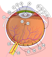
The hyaloid artery or Hyaloidarterie is an arteriole in the eye to the blood supply of the lens (11) and vitreous body (12). It is the terminal branch of the central retinal artery and runs from the exit point of the optic nerve (18) to the posterior lens pole, where it divides into the posterior part of a vascular network around the lens called the tunica vasculosa lentis . In the glass body it is of the as Gliahülle formed Canalis hyaloideus surround (13) after its Erstbeschreiber Jules Germain Cloquet also Cloquet channel is called. The hyaloid artery is normally only present during the embryonic development to supply the developing lens with nutrients and regresses shortly before birth .
On the inner side of the lens, after the regression of the hyaloid artery , an opacity known as the Mittendorf spot or hyaloid body may remain, which is generally of no clinical significance. A non-existent, incomplete or delayed regression is referred to as persistent hyaloid artery or hyaloid artery persistence and is generally considered to be a harmless anomaly in humans and animals that results in little or no visual impairment . However, it can also lead to restrictions of the field of vision and, if it continues to carry blood, to temporary bleeding into the vitreous humor. Remnants of the tunica vasculosa lentis can cause impaired visual acuity on the retina (18). Shrinkage of the hyaloid canal is a possible reason for retinal detachment .
literature
- Development of the eye and its auxiliary organs. In: Keith L. Moore, T. Vidhya N. Persaud: Embryology. Stages of development - early development - organogenesis - clinic. 5th edition. Elsevier, Urban & Fischer, Munich et al. 2007, ISBN 978-3-437-41112-0 , pp. 511-513.
- Development of the middle skin of the eye and the vessels for the eyeball. In: Walther Graumann, Dieter Sasse (Hrsg.): Compact textbook anatomy in four volumes. Volume 4: Sensory systems - skin - CNS - peripheral pathways. Schattauer, Stuttgart et al. 2005, ISBN 3-79-452064-5 , pp. 16-17.
- Pathological vitreous changes. Remnants of the hyaloid artery. In: Franz Grehn: Ophthalmology. 30th, revised and updated edition. Springer, Heidelberg 2008, ISBN 978-3-540-75264-6 , p. 262.
