Atlas (cervical vertebra)

The atlas is the first cervical vertebra. As the part of the spine closest to the skull , it carries the entire head . Because of this function, its name was borrowed from the titan Atlas of Greek mythology , who had to carry the heavens on his shoulders.
In clinical practice, the first cervical vertebra is abbreviated as C1 . In anatomy, the term atlas is used for all umbilical animals .
shape
Because of their special position and stress, the atlas and also the second cervical vertebra, axis ( abbreviated in Clinic C2 ), with which it forms a functional unit, have a different, specialized shape from the other vertebrae .
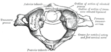
The atlas has evolutionarily lost his vertebrae - this is represented by the tooth of the Axis - and largely resembles a ring. Within this ring, on the dorsal side, the spinal cord runs from the brain and continues with its meninges in the vertebral canal that begins with the atlas through the spine.
The ring is considerably thickened on both sides slightly anteriorly . These thickenings are called massae laterales , on the upper and lower sides of which the articular surfaces to the occiput ( facies articularis superior ) and to the axis ( facies articularis inferior ) lie.
The short lateral processes, the transverse processes, lie to the side of the lateral masses . In terms of developmental history, they emerge from the costal process and have - with the exception of ruminants - the small opening foramen transversarium , which is typical of all cervical vertebrae , through which the vertebral artery runs and enters the head through the occipital opening ( foramen magnum ).
In contrast to all other vertebrae, the atlas does not have a spinous process ( processus spinosus ), but instead only a small cusp on the dorsal side of the arch ( tuberculum posterius ) (in animals tuberculum dorsale ). There is also such a small hump on the opposite front, the anterior tubercle .
Joints and ligaments
The atlas is the central element of the two head joints . It is articulated towards the skull with the occiput and below with the axis. The tooth process of the second cervical vertebra ( dens axis ) is derived ontogenetically from the vertebral body of the atlas, the intervertebral disc between the atlas and the axis as well as part of the pro- atlas and is located exactly where the vertebral body of the atlas is missing, i.e. on the ventral side of the ring between the both massae laterales . The ligamentum transversum atlantis stretches between the two massae laterales , from which longitudinal bundles ( Fasciculi longitudinales ) shear upwards to the occiput and downwards to the axis. This gives this ligament structure a cross-shaped appearance, from which the name Ligamentum cruciforme atlantis can be derived. Biomechanically, there is an extremely rare constellation here that a ligament with a real articular surface participates in a median articulatio atlantoaxialis : the front surface of the ligament contains cartilage cells and articulates with the back of the dens axis.
Developmental disorders
Due to a developmental disorder , the atlas can partially or completely grow together with the occiput, which is known as atlas assimilation . The reason for this is the complete or partial fusion of the sclerotomes from the upper four somites , from which the bony material of the vertebrae (and also of the occiput) is derived.
Injuries
The Jefferson fracture represents a special fracture of the atlas : It is a complete ring splitting of the atlas into four fracture fragments as a result of forces acting longitudinally (in the longitudinal direction of the spine). Due to the inclination of the joint surfaces on the head side, large explosive forces can be released when z. B. takes a dive into shallow water. When the neck is broken, the dens axis tears off the vertebral body of the C2 and, depending on the energy of the trauma, can also tear the cruciform ligament atlantis, thereby injuring the elongated spinal cord. If the reticular formation located here is affected, immediate death occurs with the injury. In the case of minor injuries, the dens axis can be fixed again; in such cases, access can be made orally.
literature
- G. Aumüller et al .: Anatomy. Dual series . Thieme, Stuttgart 2010, ISBN 978-3-13-136042-7 , pp. 223-226, 241.
- Jürgen Krämer, Joachim Grifka: Orthopedics, trauma surgery. Springer, Jahr, ISBN, pp. 145ff, 174f.

