Amyloid-associated angiitis

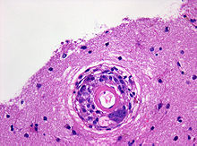
Brain biopsy with evidence of a blood vessel with a vascular wall traversed by macrophages and multinucleated giant cells. Hematoxylin-eosin stain . Original enlargement 400x

When beta-amyloid-associated angiitis (English: amyloid-beta related angiitis, ABRA ) is an inflammation of the brain supplying blood vessels (cerebral vasculitis ) with deposition of beta-amyloid called. In contrast to the common cerebral amyloid angiopathy , it is a very rare disease : fewer than 100 cases have been described so far.
The median age at diagnosis is about 67 years. The disease can manifest itself with changes in personality, headaches , seizure events or hallucinations . While magnetic resonance imaging of the skull only reveals unspecific changes in the area of the medullary bed and the lumbar puncture shows at most a moderate increase in the number of cells , the fine tissue examination of a brain tissue sample obtained from a biopsy shows granulomatous inflammatory changes and amyloid deposits in the area of small blood vessels. The lumina of the blood vessels are often thrombosed or show signs of recanalization. There are pronounced reactive changes in the surrounding brain tissue.
The clinical course and response to treatment with immunosuppressants are variable.
literature
Individual evidence
- ↑ Amick et al: Amyloid-beta-related angiitis: a rare cause of recurrent transient neurological symptoms. In: Nat Clin Pract Neurol. 2008; 4, pp. 279-283. PMID 18349854
- ↑ Scolding et al .: Abeta-related angiitis: primary angiitis of the central nervous system associated with cerebral amyloid angiopathy. In: Brain. 2005; 128, pp. 500-515. PMID 15659428 full text