Cerebral amyloid angiopathy
| Classification according to ICD-10 | |
|---|---|
| I68.0 * | Cerebral amyloid angiopathy |
| ICD-10 online (WHO version 2019) | |
The cerebral amyloid angiopathy (also cerebral amyloid angiopathy , CAA) is a disease of the blood vessels of the brain . Beta-amyloid is deposited in the walls of the blood vessels, which can narrow the lumen and lead to the formation of microaneurysms . These in turn can rupture and lead to intracerebral bleeding .
history
Cerebral amyloid angiopathy in the brains of older people was first described in 1938 by W. Scholz. Because of the commonality of amyloid deposition, CAA was originally seen as a feature of Alzheimer's disease . In the 1970s, it was possible to show that such altered vessel walls play a role in many intracerebral hemorrhages that are not due to high blood pressure.
Classification
The most common form is beta-amyloid deposition, which is seen in up to 80% of Alzheimer's disease cases. There are also various hereditary forms with mutations of the amyloid precursor protein (Dutch type), presenilin, cystatin C mutation (ACys, Icelandic type) and hereditary dementia of the British / Danish type.
Pathogenesis
Beta-amyloid is produced by cutting up the amyloid precursor protein (APP) with the help of the enzymes beta and gamma secretase . These peptides are not produced in normal metabolism ; it is assumed that the amyloid is mainly produced in nerve cells. In those affected, it accumulates in the nerve water and can be deposited in the brain tissue as so-called senile plaques , such as in Alzheimer's dementia, or in the vascular walls, as in CAA. Amyloid is mainly stored in the middle layer of the vessel wall (media) . The presence of the ApoE4 allele is associated with an increased risk of amyloid being deposited in the vessel walls.
Diagnosis
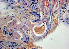
The presence of cerebral amyloid angiopathy can only be definitively confirmed by means of an autopsy or a tissue biopsy of the brain. The histology typically shows hyalinized capillaries with evidence of beta-amyloid in the vessels, often with loss of smooth muscle cells.

Histopathology of cerebral amyloid angiopathy with deposition of amyloid (red) in the wall of hyalinized meningeal vessels. Congo red staining of a brain biopsy .
|
Clinically, the diagnosis is based on the radiological presence of single or multiple lobar hemorrhages at the cortical-marrow junction or microhemorrhages in the brain with no other more likely cause. Cerebral amyloid angiopathy is considered likely if there is a history of at least two bleeds with no other apparent cause. It is estimated that between 5% and 12% of all intracranial bleeding in patients over 55 years of age is caused by cerebral amyloid angiopathy.
Individual references and sources
- ↑ W. Scholz: Studies on the pathology of the brain vessels. II The glandular degeneration of the cerebral arteries and capillaries. In: Z Neurol Psychiatr. Number 162, 1938, pp. 694-715.
- ↑ T. Revesz, JL Holton, T. Lashley, G. Plant, A. Rostagno, J. Ghiso, B. Frangione: Sporadic and familial cerebral amyloid angiopathies. In: Brain Pathol . 2002 Jul; 12 (3), pp. 343-357. Review. PMID 12146803
- ↑ a b c E. E. Smith, SM Greenberg: Beta-amyloid, blood vessels, and brain function. In: Stroke. 2009 Jul; 40 (7), pp. 2601-2606. PMID 19443808
- ^ KF Masuhr, M. Neumann: Neurology . 6th edition. Thieme, Stuttgart 2007, ISBN 978-3-13-135946-9 .
- ↑ SM Greenberg, JP Vonsattel: Diagnosis of cerebral amyloid angiopathy. Sensitivity and specificity of cortical biopsy. In: Stroke. 1997 Jul; 28 (7), pp. 1418-1422. PMID 9227694
- ^ EM Haacke, ZS DelProposto, S. Chaturvedi, V. Sehgal, M. Tenzer, J. Neelavalli, D. Kido: Imaging cerebral amyloid angiopathy with susceptibility-weighted imaging. In: AJNR Am J Neuroradiol. 2007 Feb; 28 (2), pp. 316-317. PMID 17297004

