
Coxa retrotorta
| Classification according to ICD-10 | |
|---|---|
| Q65.9 | Congenital deformity of the hip, unspecified |
| ICD-10 online (WHO version 2019) | |

Femur from the front
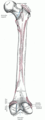
Thighbones from behind
The coxa retrotorta is the rearward rotation of the femoral neck in relation to the condylar axis, while a forward rotation represents a coxa antetorta . The normal angle of rotation varies with development, and anteversion of 10 to 20 degrees is usually present in adults. For this reason, the angle of rotation is usually specified as the angle of antetorsion, with a negative value then denoting retrotorsion.
If you look at the thigh bone from above, two lines emerge that form the anteversion angle: The base line runs through the two bony protrusions that protrude furthest backwards on the thigh condyles at knee level. The second line is the center line through the femoral neck, which is usually directed forward towards the center of the body. In the coxa retrotorta , the femoral neck axis is parallel to the baseline (anteversion angle 0 degrees) or even backwards.
A retrotorta coxa is often associated with a reduced femoral neck diaphyseal angle ( CCD angle ), i.e. a coxa vara .
The cause of the coxa retrotorta is unknown. In adults (and before the seventh embryonic month) it is within the normal range of the anteversion angle, which means that it occurs "idiopathically" in some people. In contrast to the pure coxa antetorta, however, it is considered a pre-arthrosis, i.e. a malformation that can lead to hip arthrosis at an early stage . Pain and restricted mobility usually occur in young adulthood, or earlier when the hip joint is subjected to greater physical activity. A correction can be carried out surgically in the event of relevant complaints. An intertrochanteric osteotomy is performed , whereby a simultaneous coxa vara can also be corrected.
A coxa retrotorta is more common in:
- Epiphyseolysis capitis femoris : together with a coxa vara after non-operated or incompletely corrected femoral head gliding, also after inapparent forms
- Spastic paralysis (e.g. infantile cerebral palsy ) with so-called "wind deflection deformity"
- Spina bifida with increased fixed external rotation
Measurement
The femoral neck torsion is visualized by computer tomography with incisions in the area of the femoral neck and the femoral condyles. The anteversion angle can also be displayed and measured with greater inaccuracy in special X-ray images (images according to Rippstein-Dunn, approximately also on images according to Lauenstein).
Clinical testing is also possible. The leg must be turned so that the large rolling hill can be felt as prominently as possible. In the prone position, the external rotation of the angled lower leg can be deduced directly from the anteversion, as well as in the supine position from the internal rotation of the foot with the leg extended.
Individual evidence
- ↑ S. Breusch: Clinic Guide Orthopedics Trauma Surgery. Urban & Fischer-Verlag, 2009, ISBN 978-3-437-22472-0 , p. 469. (online, with a clear sketch)
- ^ GU Exner: Normal values in pediatric orthopedics . Georg-Thieme-Verlag, Stuttgart 1990, ISBN 3-13-746301-7 .
- ↑ a b F. Hefti: Pediatric orthopedics in practice . Springer-Verlag, Berlin 1997, ISBN 3-540-61480-X .