Coxa vara
| Classification according to ICD-10 | |
|---|---|
| M21.15 | Varus deformity, not elsewhere classified -Hip joint |
| ICD-10 online (WHO version 2019) | |
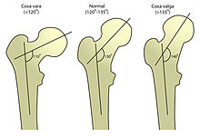
Coxa vara ( Latin coxa 'hip' , varus 'bent outwards' ) is a descriptive term for an abnormal and age-appropriate position of the upper end of the thigh bone . The (difficult to measure) CCD angle is below 126 °. If the femoral head is below the greater trochanter (CCD <90 °), one speaks of a shepherd's crook deformity.
term
Like many terms in orthopedics , “Coxa vara” is a vague term. Only the mild forms of the femoral defect are reminiscent of a "coxa vara". With Congenital coxa Vara Anglo-Saxon orthopedist denote the proximal Femurdefekt.
In individual cases, the decisive question is whether there is a malformation or a systemic disease . Rickets , fibrous dysplasia , osteogenesis imperfecta , osteodystrophia deformans and others can lead to hip varity; But this almost never has therapeutic (operative) consequences. The proximal femoral defect cannot be overlooked in children, but can be mistaken for hip dysplasia in adults .
Per- or subtrochanteric femoral fractures - however treated - can result in variations (and rotational deformities) of the upper end of the femur. Correcting them is time-consuming, complex and uncertain as a result. Endoprostheses are usually better options.
The slight varus position in intertrochanteric varus is intended. It has proven itself for decades in young adults with hip dysplasia and coxa valga .
The coxa vara always requires a (compensatory) genu valgum , the coxa valga a genu varum - and vice versa. A knee knee makes a coxa vara.
clinic
On the other hand, if the conditions are normal, functional leg shortening with limping results . The relative elevation of the greater trochanter causes the functional shortening of the gluteal splay muscles ( gluteus medius muscle ). This leads to the Trendelenburg sign and the unsteady gait of the Duchenne limp. The examination of the passive mobility while lying down shows a reduced splaying of the legs. The incentive is sometimes increased. The ability to rotate the hips should only be tested in the prone position; mostly it is laterally different and restricted in the internal rotation.
roentgen
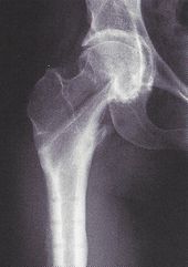
X-rays are sufficient for diagnosis and assessment of the progress. For the meaningful overview of the pelvis when standing, the feet should be closed side by side; because only then is the side comparison of the CCD angle possible. The rotation of the femoral neck is best seen on computed tomography . The femoral head pushes towards the center of the socket. This so-called centering endangers the bottom of the acetabulum . Only intertrochanteric valgization can (in healthy bones) prevent the protrusio acetabuli . This corrective osteotomy is far less useful than the intertrochanteric varus.
Developmental coxa vara
In addition to the congenital and acquired forms, a distinction is made between “extremely rare” disorders of ontogenesis (since 1928) . They are referred to as Developmental coxa vara , Infantile or cervical coxa vara or Coxa vara infantum . It develops in children between the ages of 3 and 7. Boys and girls are equally affected, in 30% on both sides. The pathogenesis is unclear. The right-angled upper end of the femur with a wide epiphyseal plate is characteristic . Although it is vertical, the femoral head has neither slipped nor rounded. The abduction osteotomy was recommended.
See also
literature
- Leonard E. Swischuk, Susan D. John: Differential Diagnosis in Pediatric Radiology . Williams & Wilkins, 1995, ISBN 0-683-08046-6 .
- Donald Resnick: Diagnosis of Bone and Joint Disorders. vol. V, Saunders, 1995, ISBN 0-7216-5071-6 .
- Fritz Hefti: Pediatric Orthopedics in Practice . Springer, 1998, ISBN 3-540-61480-X .
- Stuart Weinstein: Developmental coxa vara. In: John C. Callaghan, Aaron G. Rosenberg, Harry E. Rubash (Eds.): The Adult Hip. vol. I, Lippincott-Raven Publishers, Philadelphia / New York 1998, ISBN 0-397-51704-1 , pp. 425-427.
Individual evidence
- ↑ a b c d Stuart Weinstein: Developmental coxa vara. In: John C. Callaghan, Aaron G. Rosenberg, Harry E. Rubash (Eds.): The Adult Hip. vol. I. Lippincott-Raven Publishers, Philadelphia / New York 1998, ISBN 0-397-51704-1 , pp. 425-427.
- ↑ With the Trendelenburg sign, the healthy side sinks when standing on one leg. In the Duchenne sign, the trunk tilts to the sick side to maintain balance.
- ^ Rüdiger Döhler : Lexicon of orthopedic surgery . Springer, Berlin 2003, ISBN 3-540-41317-0 , pp. 23-24.
- ↑ J. Serafin, W. Szulc: Coxa vara infantum. In: Clinical Orthopedics and Related Research . 272, 1991, p. 103.
- ↑ CFA Bos, RJB Sakkers, JL Bloem et al: Histological, biochemical and MRI studies on the growth plate in congenital coxa vara. In: Journal of Pediatric Orthopedics. 9, 1989, p. 660.
- ^ JN Weinstein, KN Kuo, EA Miller: Congenital coxa vara. A retrospective review. In: Journal of Pediatric Orthopedics. 4, 1984, p. 70.


