
Dermatofibrosarcoma protuberans
| Classification according to ICD-10 | |
|---|---|
| C44 | Other malignant neoplasms of the skin Dermatofibrosarcoma protuberans |
| ICD-10 online (WHO version 2019) | |
The Dermatofibrosarcoma protuberans is a rare tumor of the skin, the locally aggressive, infiltrative growth, but rarely metastasizes (are given 5% to less than 0.5% of cases). After surgical therapy, the tumor tends to recur locally .
Occurrence
The tumor is rare , with an incidence of less than 1 per 100,000 population per year. It mostly affects patients around the age of 40, with men and women equally affected. The tumor is extremely rare in childhood.
Appearance
The tumor usually occurs on the trunk and on the parts of the extremities that are close to the body . It spreads irregularly in the skin and in the subcutaneous fatty tissue and has a coarse consistency. Usually it appears skin-colored. Yellow, brown or reddish discolorations are possible.
Diagnosis
A reliable diagnosis is usually only possible through a histological examination . The tumor must be differentiated from various forms of benign dermatofibroma and dermatomyofibroma on the one hand and from the prognostically much worse pleomorphic sarcoma (malignant fibrous histiocytoma) and other skin tumors as well as different forms of malignant melanoma .
Histologically, there is a diffuse spread along the septa of the subcutaneous fatty tissue, sometimes several centimeters long. The cells are densely packed, relatively uniformly spindle-shaped and more often CD34-positive .

Recurrence of dermatofibrosarcoma protuberans HE staining .
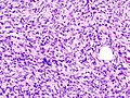
Greater magnification.

Greater magnification.
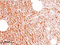
Immunohistology : CD34
_recurrence.JPG)
_recurrence.JPG)
_recurrence.JPG)
_CD34.JPG)

The preoperative examination of the tumor by sonography , computed tomography or magnetic resonance tomography can be helpful, especially in the case of deeper infiltration, but the reliable representation of the external tumor extensions is not guaranteed.
therapy
Therapy consists of surgical removal. Because the tumor can only be seen microscopically, topographical marking and complete histological processing of the excisate is recommended. If tumor extensions then extend beyond the incision margin, resection can be made at the corresponding topographical point. With this procedure, a safety distance of 1 cm is considered sufficient. Without this procedure, known as micrographic surgery , recurrences are more frequent even with significantly larger safety distances (5 cm and more).
If the tumor z. B. cannot be surgically removed due to excessive infiltration or radiation therapy may be indicated in the case of multiple recurrences .
The active ingredient imatinib , which is taken as a tablet, offers a newer, promising treatment option . Imatinib is approved for the treatment of inoperable, primary, locally recurrent, or metastatic dermatofibrosarcomata protuberans. If possible, treatment with imatinib should take place in clinical trials .
Web links
- S1 guideline for dermatofibrosarcoma protuberans of the German Dermatological Society (DDG) and the German Cancer Society. In: AWMF online (as of 2012)
- Internet portal of the German Cancer Society (DKG)
literature
- T. Mentzel, A. Beham, D. Katenkamp, AP Dei Tos, CD Fletcher: Fibrosarcomatous ("high-grade") dermatofibrosarcoma protuberans: clinicopathologic and immunohistochemical study of a series of 41 cases with emphasis on prognostic significance. In: Am J Surg Pathol . 1998, 22, pp. 576-587 PMID 9591728
- GA McArthur, GD Demetri, A. van Oosterom, among others: Molecular and clinical analysis of locally advanced dermatofibrosarcoma protuberans treated with imatinib: Imatinib Target Exploration Consortium Study B2225. In: J Clin Oncol . 2005, 23, pp. 866-873 PMID 15681532
- HM Häfner, M. Moehrle, S. Eder, B. Trilling, M. Rocken, H. Breuninger: 3D-Histological evaluation of surgery in dermatofibrosarcoma protuberans and malignant fibrous histiocytoma: Differences in growth patterns and outcome. In: Eur J Surg Oncol. 2007. (epub) PMID 17716851 .
- A. Paradis et al: Dermatofibrosarcoma protuberans: wide local excision vs. Mohs micrographic surgery. In: Cancer Treat Rev . 2008 Dec; 34 (8), pp. 728-736. Epub 2008 Aug 5. PMID 18684568