Lhermitte-Duclos syndrome
| Classification according to ICD-11 | |
|---|---|
| XH6K00 | Dysplastic gangliocytoma of cerebellum (Lhermitte-Duclos) |
| ICD-11 ( WHO version 2019) | |
The Lhermitte-Duclos disease ( LDD , including dysplastic Gangliozytom ) is a rare neoplasm of the cerebellum due to a mutation in the PTEN gene and therefore part of the PTEN tumor Hamartoma syndrome . It is also described as a subtype of Cowden syndrome .
The first description of the suffering was in 1920 by Jacques Jean Lhermitte and P. Duclos .
Pathology and histopathology

According to the WHO classification of tumors of the central nervous system, it is classified as grade I tumors. However, the syndrome shows characteristics of a benign neoplasm and a malformation at the same time, so that the exact classification is still unclear. Also, whether it could be a hamartoma is controversial in specialist circles.
The tumor suppressor gene PTEN plays a role in its development. Cowden's syndrome is based on a germline mutation in the PTEN gene. The role of the PTEN gene in the pathogenesis of Lhermitte-Duclos syndrome could be demonstrated in mouse models: A targeted elimination of PTEN in the cerebellum leads to a folding and cell migration disorder similar to the architectural disorder in humans.
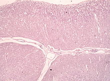
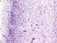
At the cellular level, one can see dysplastic (hypertrophic and later blistered, vacuolated) Purkinje cells . The stratification of the cerebellum is reversed (inverted) and the granular cell layer is largely dissolved.
Symptoms, diagnosis and therapy
Clinical symptoms and signs include headache , movement disorders (such as tremor ), visual disturbances, and an enlarged head that may mimic hydrocephalus . The symptoms increase slowly over months to years.
The electroencephalogram may be changed. The magnetic resonance imaging today provides a safe method of detection, proving the histopathological findings.
Therapy consists in the surgical removal of the tumor mass. However, recurrences are common. In some cases with familial clustering one could autosomal - dominant inheritance of the disease to be proven.
literature
- J. Lhermitte, P. Duclos: Sur un ganglioneurome diffuse du cortex du cervelet. Bulletin de l'Association francaise pour l'etude du cancer, Paris, 1920, 9: 99-107. (Initial description)
- R. Kumar, VK Vaid, SK Kalra: Lhermitte-Duclos disease. In: Child's nervous system. Volume 23, Number 7, July 2007, pp. 729-732, ISSN 0256-7040 . doi : 10.1007 / s00381-006-0271-8 . PMID 17221273 .
- N. Vantomme, F. Van Calenbergh, J. Goffin, R. Sciot, P. Demaerel, C. Plets: Lhermitte-Duclos disease is a clinical manifestation of Cowden's syndrome. In: Surgical Neurology . Volume 56, Number 3, September 2001, pp. 201-204, ISSN 0090-3019 . PMID 11597654 . (Review).
- T. Boonpipattanapong, N. Phuenpathom, W. Mitarnun: Cowden's syndrome with Lhermitte-Duclos disease. In: British journal of neurosurgery. Volume 19, Number 4, August 2005, pp. 361-365, ISSN 0268-8697 . doi : 10.1080 / 02688690500305431 . PMID 16455548 . (Review).

