
Naevus caeruleus
| Classification according to ICD-10 | |
|---|---|
| D22.L42 | Blue nevus |
| ICD-10 online (WHO version 2019) | |
A nevus caeruleus (also blue nevus or dermal melanocytoma ) is a benign, sharply delineated skin lesion that is characterized by its dark blue to gray-black color. This unusual color is caused by the proliferation of pigment-forming melanocytes in the deeper layers of the skin. It is a particular subtype of benign pigmented limited abnormalities of the skin ( pigmented nevi ), commonly known as " moles " or " liver spots ".
Epidemiology and pathogenesis
The nevus caeruleus shows no preference for gender or age; more frequent occurrences are observed towards the end of adolescence . The melanocyte precursors (melanoblasts) migrate from the neural crest into the skin during embryonic development . Melanocytes are therefore descendants of the neuroectoderm .

Typically, melanocytes should not be found in the deeper layers of the skin such as the dermis . Since this type of cell, as described, originates from the neural crest and has to migrate into the skin, it is assumed that ectopic accumulations of melanocytes can occur in the course of this process . Because of this, this lesion is sometimes called a neuron evus .
clinic
Blue nevi are most common on the back of the hand or on the dorsal side of the forearm , but they can develop anywhere. They are generally single, rough, sometimes raised nodules of blue-black color due to increased pigmentation in the dermis .
Course and prognosis
Blue nevi usually no longer change and are mainly a cosmetic problem. The transition to malignant melanoma is very rare.
In the event of sudden or unusual changes, the skin change must be excised and examined histologically in order to rule out melanoma in any case.
See also
- A → Mongolian spot is also a large-scale accumulation of deep melanocytes in the dermis, which, however, disappear again in early childhood.
- The → nevus Ota or → nevus Ito is characterized by an increase in melanocytes in the dermis of the face or shoulder that does not recede.
- Dermatofibroma
- Melanocytoma
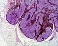
Histology of a blue nevus, Naevus coeruleus cellularis (2 ×)
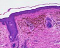
Nevus coeruleus
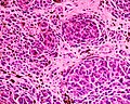
Naevus coeruleus epithelioides (10 ×)

Malignant form of a blue nevus. All histological specimens are discriminated by hematoxylin-eosin staining .


literature
- Thomas B. Fitzpatrick, Klaus Wolff (ed.): Atlas and synopsis of clinical dermatology: common and threatening diseases . 3. Edition. McGraw-Hill, New York; Frankfurt a. M. 1998, ISBN 0-07-709988-5 .
- Ernst G. Jung, Ingrid Moll (Ed.): Dermatology . 5th edition. Thieme, Stuttgart 2003, ISBN 3-13-126685-6 .
Individual evidence
- ↑ A. Osioa; M. Battistella: Le nævus bleu et ses variantes. Blue naevi and variants. In: Annales de Dermatologie et de Vénéréologie , Volume 139, October 2012, pp. 677–680