
Auricular malformation
| Classification according to ICD-10 | |
|---|---|
| Q16 | Congenital malformations of the ear that cause hearing impairment |
| Q17 | Other congenital malformations of the ear |
| ICD-10 online (WHO version 2019) | |
The term auricular malformation includes both functionally non-impairing anomalies such as protruding ears and more pronounced changes in the auricle, such as cauliflower ears , including a complete absence of the auricle. As part of the face, the auricle belongs to a prominent body region, the conspicuousness of which can be important for family life, work and social integration.
Epidemiology / etiology
Higher grade microtia occurs in 0.76 to 2.35 cases per 10,000 births in the western population. Based on the current birth rate, there are around 100 to 150 new cases per year in Germany.
Usually the malformation occurs in isolation. In 20 to 30% of cases it occurs in combination with other malformations such as facial hypoplasia , cleft lip and palate , internal malformations, cognitive impairment or as part of genetically caused syndromes , e.g. B. the auriculo-condylar syndrome or the Mengel-Konigsmark-Berlin-McKusick syndrome . The most common syndromes associated with microtia are Goldenhar syndrome and Franceschetti syndrome .
The causes are hemorrhagic events in early pregnancy , gestational diabetes and genetic causes discussed. However, further investigations are still pending. Familial clusters are observed in individual cases; this is usually based on an autosomal dominant inheritance with variable penetrance. The vast majority, however, occur sporadically. Most authors see isolated microtia as a minimal variant of hemifacial microsomia . The genesis is presumably multifactorial with an underlying genetic probability; external factors such as B. Bleeding.
Classification
When classifying auricular malformations, three grades are distinguished.
- In dysplasia ° 1 , most of the anatomical structures of the auricle are present. These include u. a. protruding ears (apostasis otum), cup-ear ° I and cup-ear ° II .
- In dysplasia ° 2 only some structures of the normal auricle are present. These include u. a. Cup ear ° III and the mini ear .
- In dysplasia ° 3 normal auricular structures are practically non-existent, only rudiments exist. These include u. a. Microtia ° III , anotia and dystopia .
Diagnosis
People with microtia must receive interdisciplinary advice and treatment at an early stage. ENT doctors, paediatricians, phoniatrists and pediatric audiologists as well as human geneticists and oral and maxillofacial surgeons should be involved. Anamestically , the family history and harmful influences in early pregnancy should be inquired about . In the case of familial accumulation, it is advisable to introduce it in human genetics in order to be able to estimate the probability of occurrence in other children. A computed tomography to assess the middle ear structures is often performed in the 10th year, usually during the first hospitalization.
Language development
According to today's doctrine, the development of spoken language should proceed undisturbed on the healthy side with a one-sided sound conduction block and normal hearing. That is why the unaffected side must be checked regularly by a specialist, and conductive or sensorineural hearing loss must be treated quickly and consistently. Children with bilateral malformations or bilateral sound conduction blocks need acoustic reinforcement soon after birth. For this purpose, the children are initially provided with a headband bone conduction hearing aid; when they are 4 years old, bone anchored devices (BAHA) can then be used. When implanting the bone anchors, care must be taken to ensure that they are not inserted too close to the rudiment of the auricle in order not to make subsequent aesthetic reconstruction more difficult with scars.
therapy
Some auricular dysplasias ° I (steel ear, cup ear, missing helix shape, saty tube, protruding auricle, curled ear (dancer type I)) should not be treated invasively even in newborns. The cartilage of the newborn is malleable in the first weeks of life and can be shaped painlessly for the newborn with an auricle former made of silicone and individually adjustable tractors. This method of treatment is most effective when started between the fifth and seventh days of the infant's life. The older the child gets, the stiffer the cartilage of the ear becomes and can at best be shaped a little. American studies show a success rate of 90% if treatment is started in the first days of life.
Surgical correction of the auricle requires at least four to five years of age in order to be physically mature for an invasive operation.
Auricular dysplasia ° I can be treated by processing existing tissue, for example by otopexy . Auricular dysplasias of a higher degree require the use of displacement plastics, transplants or implants such as the body's own cartilage or plastic. The aim should be as natural a reconstruction as possible that corresponds to the shape of the opposite side.
The surgical construction of a malformed ear with the body's own cartilage takes place in two to three steps every three months. The body's own costal cartilage is the material with which there is currently the greatest experience worldwide.
- During the first step, the rudimentary cartilage is removed, the rudimentary skin is prepared, the costal cartilage is removed and the three-dimensional auricular structure is individually manufactured and implanted from it.
- During the second step, the auricle is lifted off the ground and the fold is formed behind the ear. For this purpose, the remaining cartilage from the first step of the operation is attached behind the auricle as a spacer. The skin defect is covered both by local skin displacement and with a free skin graft.
- During the third step, corrections to the front side can be made again. The focus here is on removing excess skin from the first reconstruction step. At the same time, individual details of the auricle can be further worked out. In some cases this step can be omitted.
Alternatively, a malformed ear can be surgically rebuilt with plastic implants. The individually formable plastic implants are covered with a tissue flap from the temple area and free skin grafts. The operation can often be carried out in one step; it is not necessary to remove costal cartilage. The best long-term experience is with implants made of porous polyethylene , which have been in use since the 1980s, are manufactured industrially and are approved for surgical use. Alexander Berghaus gave the impetus for the use of polyethylene implants for ear reconstruction with experimental and clinical-scientific work.

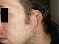
Dysplasia ° I (tail ears)
Dysplasia ° III (microtia)


Web links
Individual evidence
- ↑ Alexander Berghaus : Implants for reconstructive surgery of the nose and ear . In: Laryngo-Rhino-Otology . tape 86 , 2007, ISSN 0340-1588 , p. 67-76 , doi : 10.1055 / s-2007-966301 .