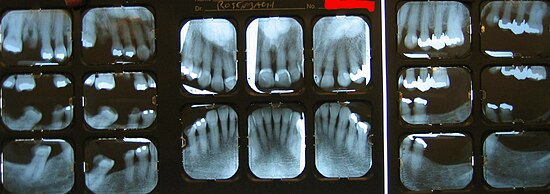
X-ray status
In dentistry, an X-ray status is the production of individual X-ray images of the entire dentition.
All tooth-bearing sections of the upper and lower jaw are represented by a series of intraoral tooth images. The recordings can be made in both analog and digital form. The advantages compared to an orthopantomogram (OPG) lie in the high level of detail recognizability of the individual images compared to the OPG, the avoidance of overprojections in the anterior tooth area, distortions and an enlargement of the image.
The X-ray status consists of 6 to 14 images. In primary teeth meet six shots in the mixed dentition ten shots. In the permanent dentition ten to fourteen shots, including so-called wing bite recordings ( ger .: Bite wing) for caries diagnosis .
Indications
An X-ray status is the method of choice in periodontal diagnostics and therapy, as it allows the periodontal bone loss in every tooth to be determined with high accuracy. This is recorded in a periodontal status. In the case of extensive prosthetic therapies , the tooth status makes its contribution to assessing the value of the pillars and the usability of teeth for supporting dentures.
Radiation exposure
In Switzerland, natural and civilization radiation leads to an annual average exposure of 4 mSv per capita. Dentistry accounts for just 0.01 mSv (1% of medical X-ray diagnostics) in the total exposure. The creation of an X-ray status using analog dental films was opposed to the higher radiation exposure compared to the OPG. The radiation exposure of a single dental film x-ray is approximately 2.1 to 5.5 µSv . With digital recording technology, the radiation exposure is only about 0.2 to 1.0 µSv, according to which the total radiation exposure of all individual images of an X-ray status is slightly lower than that of an OPG with about 19 µSv. Even if the effective dose in the 14-image dental film status and its share in the total exposure is very low, it must be taken into account that relatively high local doses (especially on the skin) are applied, which are masked by the calculation of the effective doses.
For comparison: When traveling by air at an altitude of 10 to 12 km, the radiation exposure from cosmic rays is around 5.5 µSv per hour.
Possible radiation effects
Meningiomas are benign brain tumors with an incidence of 6–8 per 100,000. According to a study by Yale University , frequent x-rays of the teeth increases the risk of developing a benign brain tumor. People who are x-rayed at the dentist's one or more times a year are three times more likely to develop meningioma. For children under ten years of age who are often x-rayed, the risk is up to five times higher.
execution
The X-ray film, or in the case of digital recording, the sensor (usually 2 × 3 cm or 3 × 4 cm) is placed in the mouth and kept as depressurized as possible by the patient himself. This process is repeated for each individual tooth film recording. Alternatively, an image receiver holder is used, which ensures the central beam guidance. This, in turn, is of great importance for an isometric representation of the tooth.
Reward
In the evaluation standard dental services (BEMA) in Germany is the reward of the x-status (more than 8 single shots) for insured patients after BEMA no. Ä925d with 34 points (around € 31). According to the private fee schedule for dentists (GOZ), each individual exposure is paid for when an X-ray status is prepared according to the GOZ no. 5000 (1.8x) at € 5.24, which results in € 52.40 for 10 exposures
Individual evidence
- ↑ Friedrich Anton Pasler: Pocket Atlas of Dental Radiology . Georg Thieme Verlag, 2003, ISBN 978-3-13-128991-9 , pp. 64f.
- ↑ A. Aroua, B. Burnand et al. a .: Nation-wide survey on radiation doses in diagnostic and interventional radiology in Switzerland in 1998. In: Health physics. Volume 83, Number 1, July 2002, pp. 46-55, ISSN 0017-9078 . PMID 12075683 .
- ↑ Michael Hülsmann: Endodontics . Georg Thieme Verlag, 8 October 2008, ISBN 978-3-13-156581-5 , p. 85–.
- ↑ J. Th. Lambrecht, Radiation exposure from analog and digital racks and panoramic film recordings Switzerland. Monthly Zahnmed 114: 687-693 (2004)
- ↑ Claus Grupen: Basic course on radiation protection . Springer-Verlag, 2008, ISBN 978-3-540-75849-5 , pp. 176–.
- ↑ Elizabeth B. Claus, Lisa Calvocoressi a. a .: Dental x-rays and risk of meningioma. In: Cancer. 118, 2012, p. 4530, doi : 10.1002 / cncr.26625 .

