Celiac trunk compression syndrome
| Classification according to ICD-10 | |
|---|---|
| I77.4 | Coeliac Artery Compression Syndrome |
| ICD-10 online (WHO version 2019) | |
The median arcuate ligament syndrome , also known as Harjola-Marable syndrome , Dunbar syndrome or ligament-arcuatum syndrome called, manifested by abdominal pain and by clamping the celiac artery ( celiac ) and possibly the celiac ganglion through the diaphragm causes . The abdominal pain may be related to food intake and may be accompanied by weight loss. During the auscultation of the abdomen, one often hears typical stenosis noises .
The diagnosis of celiac trunk compression syndrome is a diagnosis of exclusion because many people have this form of entrapment without developing symptoms. Therefore, the diagnosis can only be made after other causes have been ruled out. Duplex sonography is used for screening ; the diagnosis is confirmed using computed tomography (CT) or magnetic resonance imaging (MRT).
Treatment is surgically. The ligament is severed and the aortic hiatus is expanded. The celiac ganglion may also be removed. The majority of patients benefit from the operation. The success rate is lower in younger patients, patients with psychiatric illnesses, patients with increased alcohol consumption, patients without weight loss, and patients who do not experience pain related to meals.
Anatomy and pathogenesis
The ligamentum arcuatum medianum arises at the base of the diaphragm, where the right and left diaphragmatic crus (crus dextrum et sinistrum) come together approximately at the level of the 12th thoracic vertebra. This tissue arch forms the front of the aortic hiatus, through which the aorta with the aortic plexus and the thoracic duct pass. Usually the ligament sits above the origin of the celiac trunk, but in about 25% of people the ligament crosses at the level of the origin and thereby constricts the artery and adjacent structures such as the celiac ganglion. In some, the constriction is so severe that the symptoms of the disease arise.
Various theories try to explain the pain caused by compression. One suspects that the cause of the pain is insufficient blood flow ( ischemia ) in the abdominal organs supplied, another assumes that the celiac ganglion is compressed.
history
A compression of the trunk was first observed by Benjamin Lipshutz in 1917. Celiac trunk compression syndrome was described in 1963 by Pekka-Tapani Harjola and two years later by J. David Dunbar and Samuel Marable.
Epidemiology
Only about 1% of people in whom the ligament crosses at the level of the trunk will have celiac trunk compression syndrome. The complaints mainly affect patients between the ages of 20 and 40, mostly women, preferably of a smaller build.
Complaint picture
The affected people complain of nausea and burning, cramp-like pain, which are often located in the epigastrium and not infrequently occur in connection with meals. The pain can lead to weight loss and even anorexia . Occasionally a sound of stenosis in the epigastrium can be auscultated . Complications arise from compression of the artery, such as: B. gastric paralysis or aneurysmal enlargement of the superior pancreaticoduodenal artery , which serves as a collateral because of its connection via the inferior pancreaticoduodenal artery to the superior mesenteric artery .
Diagnosis
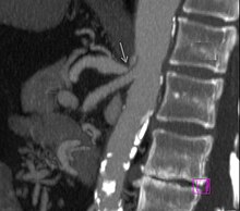
| CT angiographic findings in celiac trunk compression syndrome |
|---|
|
The diagnosis of exclusion includes esophago-gastro-duodenoscopy and colonoscopy . Gall complaints and reflux esophagitis must also be excluded. The diagnosis is ultimately based on the combination of the symptoms with the radiological diagnosis. The classic triad abdominal pain-weight loss-stenosis noise is only found in a few patients. Radiological diagnostics are divided into:
- Screening: duplex sonography to measure the blood flow in the celiac trunk. A blood flow velocity of> 200 cm / s is considered suspicious
- Diagnostics: In the past, angiography was performed to confirm the diagnosis , which has now been replaced by CT angiography or MRT angiography , with the CT examination being preferred because of the better representation of the neighboring abdominal organs.

The findings of a short narrowing of the celiac trunk at its outlet with subsequent expansion (poststenotic dilatation), a notch in the upper aspect of the trunk and a hook-shaped course of the trunk support the diagnosis of a celiac trunk compression syndrome. These image criteria are emphasized in expiration and are sometimes even found in asymptomatic patients who do not suffer from the syndrome.
Other possible differential diagnoses in the case of a narrowing near the outlet with poststenotic dilatation, such as arteriosclerotic changes , must also be taken into account. Here, the hook-shaped course of the celiac trunk can be helpful for differentiation, although this criterion is also not pathognomonic for celiac trunk compression syndrome. The frequency for this anatomy in normal asymptomatic individuals is given as 10 to 24%.
therapy
Decompression of the trunk is the therapy of choice. This is usually done through a laparotomy with the aim of detaching the ligament from the artery. At the same time, the celiac ganglion is removed and the blood flow of the freed artery is checked using duplex sonography. If the reduced blood flow persists, revascularization through a bypass or other vascular surgical interventions may be necessary.
Decompression can also take place via a laparoscopic approach, but if revascularization of the trunk is necessary, the open approach must be changed.
Endoscopic procedures such as percutaneous transluminal angioplasty (PTA) have been used in patients who have not been able to access open or laparoscopic access, where PTA alone, without decompression of the artery by the ligament, has not been successful.
forecast
There are few studies of long-term outcomes from the treatment of patients with celiac compression syndrome. Duncan's paper reports on a study of 51 patients who were operated on through an open approach. 44 of these patients could be followed up over a period of nine years. 75% of the patients who underwent decompression as well as revascularization remained symptom-free. The following were named as predictors of good success:
- Age between 40 and 60 years
- no psychiatric abnormalities and alcohol abstinence
- weight loss suffered> 9 kg
A more recent study from 2009 also indicates the success rate for surgical therapy at around 70–75%.
Individual evidence
- ↑ a b c d e f g h i j k K. M. Horton, MA Talamini, EK Fishman: Median arcuate ligament syndrome: evaluation with CT angiography . In: Radiographics . tape 25 , no. 5 , 2005, p. 1177-1182 , doi : 10.1148 / rg.255055001 , PMID 16160104 .
- ^ A b B. Luther: Celiac trunk compression syndrome . In: Wolfgang Hepp, Helmut Kogel (Ed.): Vascular surgery . Elsevier, Urban & Fischer, Munich / Jena, ISBN 3-437-21841-7 .
- ↑ HH Lindner, E. Kemprud: A clinicoanatomical study of the arcuate ligament of the diaphragm . In: Arch Surg . tape 103 , no. 5 , November 1971, p. 600-605 , PMID 5117015 .
- ↑ a b c d e f g h i A. A. Duncan: Median arcuate ligament syndrome . In: Curr Treat Options Cardiovasc Med . tape 10 , no. 2 , April 2008, p. 112-116 , doi : 10.1007 / s11936-008-0012-2 , PMID 18325313 .
- ^ PT Harjola: A rare obstruction of the coeliac artery. Report of a case . In: Ann Chir Gynaecol Fenn . tape 52 , 1963, pp. 547-550 , PMID 14083857 .
- ^ JD Dunbar, W. Molnar, FF Beman, SA Marable: Compression of the celiac trunk and abdominal angina . In: Am J Roentgenol Radium Ther Nucl Med . tape 95 , no. 3 , November 1965, p. 731-744 , PMID 5844938 ( ajronline.org ).
- ↑ DH Balaban, J. Chen, Z. Lin, CG Tribble, RW McCallum: Median arcuate ligament syndrome: a possible cause of idiopathic gastroparesis . In: Am. J. Gastroenterol. tape 92 , no. 3 , March 1997, p. 519-523 , PMID 9068484 .
- ↑ NE Manghat, G. Mitchell, CS Hay, IP Wells: The median arcuate ligament syndrome revisited by CT angiography and the use of ECG gating - a single center case series and literature review . In: Br J Radiol . tape 81 , no. 969 , September 2008, p. 735-742 , doi : 10.1259 / bjr / 43571095 , PMID 18541631 .
- ^ IA Sproat, MA Pozniak, TW Kennell: US case of the day. Median arcuate ligament syndrome (celiac artery compression syndrome) . In: Radiographics . tape 13 , no. 6 , November 1993, pp. 1400-1402 , PMID 8290734 .
- ↑ a b c D. Grotemeyer, M. Duran, F. Iskandar, D. Blondin, K. Nguyen, W. Sandmann: Median arcuate ligament syndrome: vascular surgical therapy and follow-up of 18 patients . In: Langenbecks Arch Surg . tape 394 , no. 6 . Springer-Verlag, 2009, p. 1085-1092 , doi : 10.1007 / s00423-009-0509-5 , PMID 19506899 .
- ↑ AM Carbonell, KW Kercher, BT Heniford, BD Matthews: Multimedia article. Laparoscopic management of median arcuate ligament syndrome . In: Surg Endosc . tape 19 , no. 5 , May 2005, pp. 729 , doi : 10.1007 / s00464-004-6010-x , PMID 15965588 .
- ↑ AH Matsumoto, CJ Tegtmeyer, EK Fitzcharles et al: Percutaneous transluminal angioplasty of visceral arterial stenoses: results and long-term clinical follow-up . In: J Vasc Interv Radiol . tape 6 , no. 2 , 1995, p. 165-174 , doi : 10.1016 / S1051-0443 (95) 71087-9 , PMID 7787348 .
- ^ LM Reilly, AD Ammar, RJ Stoney, WK Ehrenfeld: Late results following operative repair for celiac artery compression syndrome . In: J. Vasc. Surg. tape 2 , no. 1 , January 1985, p. 79-91 , PMID 3965762 ( full text of the article ).

