
Notochord: Difference between revisions
No edit summary |
|||
| Line 18: | Line 18: | ||
DorlandsSuf = 12579301 | |
DorlandsSuf = 12579301 | |
||
}} |
}} |
||
The '''notochord''' is a flexible, rod-shaped body found in [[embryo]]s of all [[chordate]]s. It is composed of [[cell (biology)|cell]]s derived from the [[mesoderm]] and defines the primitive axis of the [[embryo]]. In some [[vertebrate]]s and [[chordate]]s, it persists throughout life as the main [[axial skeleton|axial support]] of the body, while in most vertebrates it is replaced by the [[vertebral column]]. The notochord is found [[anatomical terms of location|ventral]] to the [[neural tube]]. |
The '''notochord''' is a flexible, dick rod-shaped body found in [[embryo]]s of all [[chordate]]s. It is composed of [[cell (biology)|cell]]s derived from the [[mesoderm]] and defines the primitive axis of the [[embryo]]. In some [[vertebrate]]s and [[chordate]]s, it persists throughout life as the main [[axial skeleton|axial support]] of the body, while in most vertebrates it is replaced by the [[vertebral column]]. The notochord is found [[anatomical terms of location|ventral]] to the [[neural tube]]. |
||
Notochords were the first "backbones" serving as support structures in chordates such as ''[[Haikouicthys]]''. Notochords were advantageous to primitive fish-ancestors because they were a rigid structure for muscle attachment, yet flexible enough to allow more movement than, for example, the exoskeleton of the dominant animals of that time. Embryos of vertebrates have notochords today, as embryonic development often happens to follow a pattern similar to the ancestral evolution of the modern animal's traits. In [[tetrapod]]s, they eventually develop into the [[nucleus pulposus]] of the [[intervertebral discs]]. |
Notochords were the first "backbones" serving as support structures in chordates such as ''[[Haikouicthys]]''. Notochords were advantageous to primitive fish-ancestors because they were a rigid structure for muscle attachment, yet flexible enough to allow more movement than, for example, the exoskeleton of the dominant animals of that time. Embryos of vertebrates have notochords today, as embryonic development often happens to follow a pattern similar to the ancestral evolution of the modern animal's traits. In [[tetrapod]]s, they eventually develop into the [[nucleus pulposus]] of the [[intervertebral discs]]. |
||
Revision as of 01:43, 24 June 2009
| Notochord | |
|---|---|
 Transverse section of a chick embryo of forty-five hours’ incubation. | |
| Details | |
| Precursor | chordamesoderm |
| Gives rise to | nucleus pulposus |
| Identifiers | |
| MeSH | C000000 |
| TE | E5.0.1.1.0.0.8 |
| FMA | 85521 |
| Anatomical terminology | |
The notochord is a flexible, dick rod-shaped body found in embryos of all chordates. It is composed of cells derived from the mesoderm and defines the primitive axis of the embryo. In some vertebrates and chordates, it persists throughout life as the main axial support of the body, while in most vertebrates it is replaced by the vertebral column. The notochord is found ventral to the neural tube.
Notochords were the first "backbones" serving as support structures in chordates such as Haikouicthys. Notochords were advantageous to primitive fish-ancestors because they were a rigid structure for muscle attachment, yet flexible enough to allow more movement than, for example, the exoskeleton of the dominant animals of that time. Embryos of vertebrates have notochords today, as embryonic development often happens to follow a pattern similar to the ancestral evolution of the modern animal's traits. In tetrapods, they eventually develop into the nucleus pulposus of the intervertebral discs.
Development of the notochord
Notogenesis is the development of the notochord by the epiblasts that make up the floor of the amnion cavity (Human Embryology). The notochord arises as a pouch from the mesoderm.
The notochord forms during gastrulation and soon after induces the formation of the neural plate (neurulation), synchronizing the development of the neural tube. On the ventral aspect of the neural groove an axial thickening of the endoderm takes place. (In bi-pedal chordates, e.g. humans, this surface is properly referred to as the anterior surface). This thickening appears as a furrow (the chordal furrow) the margins of which anastomose (come into contact), and so convert it into a solid rod of polygonal-shaped cells (the notochord) which is then separated from the endoderm.
It extends throughout the entire length of the future vertebral column, and reaches as far as the anterior end of the midbrain, where it ends in a hook-like extremity in the region of the future dorsum sellæ of the sphenoid bone. Initially it exists between the neural tube and the endoderm of the yolk-sac, but soon becomes separated from them by the mesoderm, which grows medially and surrounds it. From the mesoderm surrounding the neural tube and notochord, the skull, vertebral column, and the membranes of the brain and medulla spinalis are developed.
Postembryonic vestige of the notochord is found in the nucleus pulposus of the intervertebral disks, but not in the vertebral bodies, from which notochordal cells usually regress entirely. In humans, by the age of 4, all notochord residue is replaced by a population of chondrocyte-like cells of unclear origin[1]. Persistence of notochordal cells within the vertebra may cause a pathologic condition- persistent notochordal canal[2]. They are also found to persist in the nasopharyngeal space and, in such an unusual instance, may give rise to Tornwaldt's cyst.
The notochord in neural development
Research into the notochord has played a key role in understanding the development of the central nervous system. By transplanting and expressing a second notochord near the dorsal neural tube, 180 degrees opposite of the normal notochord location, one can induce the formation of motoneurons in the dorsal tube. Motoneuron formation generally occurs in the ventral neural tube, while the dorsal tube generally forms sensory cells.
The notochord secretes a protein called sonic hedgehog homolog (SHH), a key morphogen regulating organogenesis and having a critical role in signaling the development of motoneurons[3]. The secretion of SHH by the notochord establishes the ventral pole of the dorsal-ventral axis in the developing embryo.
The structure of the notochord
The notochord is composed primarily of a core of glycoproteins, encased in a sheath of collagen fibers wound into two opposing helices. The angle between these fibers determines whether increases pressure in the core will result in shortening and thickening versus lengthening and thinning.
Organisms with a notochord
Additional images
-
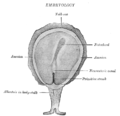 Surface view of embryo of Concolor gibbon (Hylobates concolor).
 -
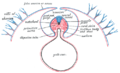 Diagram of a transverse section, showing the mode of formation of the amnion in the chick.
 -
 Section through the head of a human embryo, about twelve days old, in the region of the hind-brain.
 -
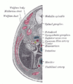 Transverse section of human embryo eight and a half to nine weeks old.


References
- ^ Urban, J. P. G. (2000). "The Nucleus of the Intervertebral Disc from Development to Degeneration". Integrative and Comparative Biology. 40: 53. doi:10.1093/icb/40.1.53.
- ^ Christopherson, Lr; Rabin, Bm; Hallam, Dk; Russell, Ej (1999). "Persistence of the notochordal canal: MR and plain film appearance" (Free full text). AJNR. American journal of neuroradiology. 20 (1): 33–6. ISSN 0195-6108. PMID 9974055.
{{cite journal}}: Unknown parameter|day=ignored (help); Unknown parameter|month=ignored (help)CS1 maint: multiple names: authors list (link) - ^ Echelard, Y; Epstein, Dj; St-Jacques, B; Shen, L; Mohler, J; Mcmahon, Ja; Mcmahon, Ap (1993). "Sonic hedgehog, a member of a family of putative signaling molecules, is implicated in the regulation of CNS polarity". Cell. 75 (7): 1417–30. doi:10.1016/0092-8674(93)90627-3. PMID 7916661.
{{cite journal}}: Unknown parameter|month=ignored (help)CS1 maint: multiple names: authors list (link)