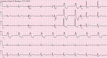
Accelerated idioventricular rhythm
The accelerated idioventricular rhythm (AIVR) is an ectopic ventricular rhythm of at least 3 consecutive beats with a rate between 50 and 120 beats per minute. Since the origin of the cardiac excitation lies in the ventricle (bundle of His, Purkinje system or working myocardium), the ECG shows a characteristically widened QRS complex . The AIVR is usually benign and well tolerated.
Pathophysiology
Normally, ventricular (so-called tertiary) pacemaker centers only come into action when the sinus rhythm fails or there is a conduction blockage ( AV block ). In the case of the AIVR, however, a ventricular pacemaker center shows pathologically increased activity. The ectopic pacemaker center then takes over the pacemaker function of the heart as soon as its frequency is above the frequency of the sinus node.
AIVR often occurs in the reperfusion phase after an acute myocardial infarction and is therefore a non-specific marker for the recirculation of blood in the artery that is blocked during a myocardial infarction. Whether the AIVR indicates that the infarct artery is fully open again is a matter of controversy. Other causes can be cocaine or digitalis poisoning or an inflammation of the myocardium ; it also occurs after cardiac surgery.
The AIVR can also occur in people with healthy hearts, especially athletes. In this case, increased vagal tone is the cause. This leads to a very slow sinus rhythm which ventricular pacemaker centers can easily overtake.
ECG criteria and differentiation from other arrhythmias
The beginning and the end of the AIVR is generally slow, the frequency is between 40-50 and 120 beats per minute, depending on the literature. It is largely regular, usually short-lived and self-limiting. The chamber complex is branch block-like deformed, in distinction from a sinus rhythm but exists with bundle branch block is no temporal correlation between P-wave ( atrial excitation ), and QRS complex (ventricular excitation). A Ventricular tachycardia (VT) will generally have a higher frequency (> 120 / min), but the definition of a slow VT can be difficult. A ventricular reserve rhythm can be assumed at a lower rate (<40–50 / min).
Therapy and prognosis
Since the arrhythmia is usually self-limiting and hemodynamically irrelevant, therapy is usually not required. In the case of hemodynamic impairment, it is recommended e.g. B. to raise the base frequency of the sinus node by atropine so that it takes over the pacemaker function again, and not to suppress the idioventricular rhythm. Stimulation by a pacemaker in the atrium is also possible.
The AIVR has a good prognosis for people with healthy hearts and athletes. In patients with a heart attack who immediately received interventional therapy using a cardiac catheter, the occurrence of an AIVR was not associated with either an improved or a worsened prognosis. AIVR can be particularly problematic in patients with a right heart attack when the right coronary artery is blocked , since bradycardia and haemodynamic consequences are more likely than in other situations.
Individual evidence
- ↑ a b c d H. Bonnemeier, J. Ortak, UK Wiegand, F. Eberhardt, F. Bode, H. Schunkert, HA Katus, G. Richardt: Accelerated idioventricular rhythm in the post-thrombolytic era: incidence, prognostic implications, and modulating mechanisms after direct percutaneous coronary intervention. In: Annals of noninvasive electrocardiology: the official journal of the International Society for Holter and Noninvasive Electrocardiology, Inc. Volume 10, Number 2, April 2005, ISSN 1082-720X , pp. 179-187, doi: 10.1111 / j.1542- 474X.2005.05624.x , PMID 15842430 .
- ↑ a b c d A. R. Riera, RB Barros, FD de Sousa, A. Baranchuk: Accelerated idioventricular rhythm: history and chronology of the main discoveries. In: Indian pacing and electrophysiology journal. Volume 10, Number 1, 2010, ISSN 0972-6292 , pp. 40-48, PMID 20084194 , PMC 2803604 (free full text).
- ↑ Gerd Herold : Internal Medicine - A lecture-oriented presentation. Herold Verlag, Cologne 2011. p. 270
- ↑ a b c d e f Fauci et al (Ed.): Harrison's internal medicine . 17th edition. ABW, Berlin 2009, ISBN 978-3-86541-310-9 , pp. 1769 .
- ↑ Glaser F., Rohla M .: EKG differential diagnosis of the wide-QRS complex tachycardia In: Journal für Kardiologie - Austrian Journal of Cardiology 2008; 15 (7-8), 218-235
