Antagonist (muscle)
The antagonist is a muscle and an opponent of the agonist . The muscular interaction of the limbs of the body is also known as the antagonist principle.
Classical anatomy assumes that this principle can be described using the example of the flexor and extensor muscles on the arm as follows: If the flexor ( biceps ) is actively shortened when the arm is bent, the extensor ( triceps ) is passively stretched at the same time. Conversely, if the extensor is actively shortened while extending the arm, the flexor is passively stretched.
In fact, almost all of the antagonists contribute to the movement and are also innervated ( tone ) at rest . In many cases (e.g. shoulder joint) a movement would not be possible without dislocating the joint if the antagonist did not contract in a certain ratio to the agonist. This is an eccentric contraction .
There are also numerous muscles that are formally regarded as antagonists but act as synergists . This is especially the case with muscles stretching across two joints. A well-known example are the muscles of the so-called ischiocrural muscles , which are involved in stretching movements of the knee (getting up from a seat, starting a sprint, squatting, etc.), although they are assigned a flexing function in relation to the knee joint ( Lombard ' paradox ).
Example eye muscles
The antagonistic principle of action can also be observed in the pupillary reaction, in which the expansion and constriction occur through the two inner eye muscles, the dilator pupillae and the sphincter pupillae muscles .
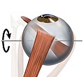
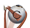
Each human eye also has six outer eye muscles that are responsible for its coordinated movements. Two of them each have a similar muscle level and turn the eye around an almost identical axis of rotation, but each in an opposite direction of rotation. These muscles are called antagonists. In contrast, muscles that move the eye around a similar axis of rotation in the same direction are called synergists . This terminology is also used when only partial functions of the respective muscles agree or counteract one another. It is only fully applicable for ductions, i.e. movements of one eye. If one extends the consideration to the opposite eye, this definition for vergences, opposing eye movements, has to be restricted in order to describe contralateral synergists and antagonists.
The movements of the eyes are finally carried out by a reciprocal change in the innervation. For example, Sherrington's law states that the innervation of an antagonist decreases as that of the agonist increases. The fact that this also applies equally to the contralateral synergists and antagonists of the other eye, says Hering's law of the same side innervation .
- Ipsilateral (equilateral) synergists and antagonists with regard to the respective muscle function
| Agonist | function | Synergists | Antagonists |
|---|---|---|---|
| M. rect. Medialis | Adduction |
Rect. Superior, rect. Inferior |
M. rect.lateralis, M. obl. Superior, M. obl. Inferior |
| Lateral rectus muscle | Abduction |
M. obl. Superior, M. obl. Inferior |
M. rect.medialis, M. rect. Superior, M. rect. Inferior |
| M. rect. Superior | Elevation
Inside roll Adduction |
M. obl. Inferior
M. obl. Superior M. rect.medialis, M. rect.inferior |
M. rect.inferior, M. obl. Superior
M. rect. Inferior, M. obl. Inferior M. rect.lateralis, M. obl. Superior, M. obl. Inferior |
| M. rect. Inferior | Lowering
Outside roll Adduction |
M. obl. Superior
M. obl. Inferior Medial rectus muscle, superior rectal muscle |
M. rect. Superior, M. obl. Inferior
M. obl. Superior, M. rect. Superior M. rect.lateralis, M. obl. Superior, M. obl. Inferior |
| M. obl. Superior | Lowering
Inside roll Abduction |
M. rect. Inferior
M. rect. Superior M. rect.lateralis, M. obl. Inferior |
M. rect. Superior, M. obl. Inferior
M. rect. Inferior, M. obl. Inferior M. rect.medialis, M. rect. Superior, M. rect. Inferior |
| M. obl. Inferior | Elevation
Outside roll Abduction |
M. rect. Superior
M. rect. Inferior M. rect.lateralis, M. obl. Superior |
M. rect.inferior, M. obl. Superior
M. obl. Superior, M. rect. Superior M. rect.medialis, M. rect. Superior, M. rect. Inferior |
- Graphic representation of the involvement of individual muscles (synergists) in the respective rotational movements using the example of the right eye
| Elevation. The main participants are the M. rectus superior and the M. obliquus inferior | Lowering. The main participants are the M. rectus inferior and M. obliquus superior | Adduction. The main involved is the M. rectus medialis and the vertical Mm. recti | Abduction. The main involved is the lateral rectus muscle and the Mm. obliqui | Inside roll. The main participants are the M. obliquus superior and the M. rectus superior | Outside roll. The main participants are the M. obliquus inferior and the M. rectus inferior |
literature
- Lexicon of Biology. 1st volume, Spektrum Akademischer Verlag, Heidelberg 2004, ISBN 3-8274-0326-X .
- Th. Axenfeld (conception), H. Pau (ed.): Textbook and atlas of ophthalmology . With the collaboration of R. Sachsenweger and others Gustav Fischer Verlag, Stuttgart 1980, ISBN 3-437-00255-4 .
- Albert J. Augustin: Ophthalmology . Springer Verlag, Berlin 2007, ISBN 978-3-540-30454-8 .
Individual evidence
- ^ Herbert Kaufmann: Strabismus. With the collaboration of W. de Decker et al. Enke, Stuttgart 1986, ISBN 3-432-95391-7 , p. 38.
- ^ Herbert Kaufmann : Strabismus. With the collaboration of W. de Decker et al. Enke, Stuttgart 1986, ISBN 3-432-95391-7 , p. 61 ff.
- ^ Albert J. Augustin: Ophthalmology . Springer-Verlag, 2007, ISBN 978-3-540-30454-8 .


