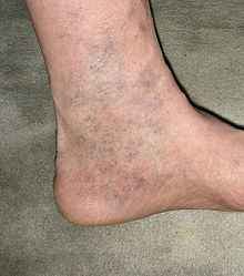
Spider veins
Spider veins (in the jargon of the internal medicine as a special subtype of varicosis defined, therefore, spider veins called) are small, directly (intradermal) in the epidermis located and clearly visible mesh or fan-shaped veins . Over 60% of the population are affected. Together with the reticular varices, which have a larger diameter of up to 3 mm and lie somewhat deeper in the skin, doctors group them together as so-called C1 varices (according to the CEAP classification ).
Spider veins mainly appear on the legs. They can be the result of a congenital predisposition or a congestion in the venous system. In this case, the fine veins lose their original elasticity over time due to permanently increased pressure, they widen and become visible as red-bluish tortuous vascular structures. Increasing age, pregnancy, hormone preparations such as birth control pills, lack of exercise, standing jobs and being overweight can encourage the development of spider veins or worsen the symptoms.
Although spider veins without involvement of the rest of the leg vein system are not a disease in the medical sense, they represent a significant cosmetic problem for many people. However, spider veins can also be a first indication of a disease of the deeper venous system.
treatment
In a study it could be shown that spider veins like larger varicose veins no longer have properly closing (micro) venous valves. From this it can be concluded that spider veins also represent pathological changes in the venous vascular system. For this reason, the entire superficial and deep venous system should ideally be examined prior to treatment in order to rule out larger varicose veins , leading venous insufficiency or chronic venous insufficiency (CVI). The examination is carried out using a non-invasive and painless ultrasound examination. If a pathological change in the leg vein system is found in addition to the spider veins or as their cause, this must be treated accordingly.
Spider veins can be treated with sclerotherapy (sclerotherapy) and very fine spider veins can also be treated with various transdermal lasers. Sclerotherapy is recommended as the therapy of choice for spider veins according to current medical guidelines. With this treatment method, a suitable sclerosant (the active ingredient polidocanol or macrogollauryl ether is approved in Germany ) is slowly injected directly into the spider veins with a very fine needle. For spider veins and their nutrient veins, Polidocanol in concentrations 0.25; 0.5 or 1% applied. The sclerosant leads to targeted and desired damage to the vein wall. This leads to the activation of the body's own signaling pathways, which over time cause the damaged vein to transform into a connective tissue cord. This process is also known as sclerosis and the therapy is therefore also known as sclerotherapy or sclerotherapy. The connective tissue strands are then broken down by the body in the long term. A discoloration of the vein immediately after the injection started indicates that the sclerosant has displaced the blood and that the injection was made intravascularly. The remodeling process described above takes some time so that the success of the treatment may only be visible after a few weeks.
Reticular varices often represent nutrient veins of spider veins. These nutrient veins supply the spider veins with blood and should therefore be treated to get rid of the spider veins. An untreated nutritional vein is often the reason for spider veins to recur in the treated area. Due to the shallow penetration depth, nutrient veins can usually not be removed by transdermal lasers.
Instead of a polidocanol solution, foamed polidocanol (microfoam) can also be used. The foam is also injected into the spider veins with the finest needles and is considered an additional treatment option for C1 varices. Foamed polidocanol has the advantage over the liquid sclerosant that the blood is better displaced in the veins to be treated and longer contact of the polidocanol with the vein wall increases the sclerosis effect. However, this can also increase the risk of undesirable effects. Foam sclerotherapy is therefore preferably used to treat larger varicose veins (reticular varices, lateral branch and trunk varices).
The sclerosing fluid is broken down and excreted by the body within 48 hours. The treatment usually lasts 15–20 minutes. The result can be optimized through repeated sessions and an improvement of over 90% can be achieved. If carried out properly, treatment with Polidocanol is an efficient form of therapy with few side effects. Side effects such as temporary brown discoloration of the skin ( hyperpigmentation ) or new formation of small vessels in the course of the treated varices ( called matting ) can occur. Compression following the injection and wearing it consistently for one to three weeks can optimize the result.
Isolated spider veins (up to 0.1 mm in diameter) lying on the very surface can also be treated with special lasers. The health insurance usually does not cover the treatment, as it is a purely cosmetic problem.
Word origin
The word component Reiser comes from botany , where rice means as much as twig (see sticks ); The spider veins got their name due to the external similarity of the veins to the thin twigs that were processed into (sweeping) brooms until the last century.
Web links
Individual evidence
- ^ Rabe E, Pannier-Fischer F, Bromen K, Schuldt K, Stang A, Poncar CH, Wittenhorst M, Bock E, Weber S, Jöckel KH: Bonner Venenstudie der Deutsche Gesellschaft für Phlebologie In: Phlebologie 32, 2003, p. 1 -14. doi: 10.1055 / s-0037-1617353
- ^ Wienert V, Simon HP, Böhler U: Angioarchitecture of spider veins. Scanning electron microscope study of corrosion specimens In: Phlebologie 35, 2006, pp. 24-29. doi: 10.1055 / s-0037-1622127
- ↑ a b c Erika Mendoza: Guide to varicose veins, leg swelling and thrombosis . Springer Verlag, Heidelberg 2016, ISBN 978-3-662-49738-8 , pp. 186-188 .
- ↑ a b c d Rabe E, Breu FX, Flessenkämper I, Gerlach H, Guggenbichler S, Kahle B, Murena R, Reich-Schupke S, Schwarz T, Stücker M, Valesky E, Werth S, Pannier F: Guideline: Sclerotherapy treatment of the Varicose veins. AWMF guideline no. 037-015, status 12-2018, accessed on March 10, 2020
- ↑ Eurocom eV (Ed.): "Vein diseases and their therapy", 3rd edition, Düren. P. 33. 2012
- ^ Eberhard Rabe, Markus Stücker Ed .: Phlebological picture atlas . Viavital Verlag, Cologne 2013, ISBN 978-3-934371-52-1 , p. 269 .
- ↑ Kahle B, Leng K: Efficacy of sclerotherapy in varicose veins - a prospective, blinded placebocontrolled study In: Dermatol. Surg 30, 2004, pp. 723-728. doi: 10.1111 / j.1524-4725.2004.30207.x
- ↑ Rabe E, Schliesshake D, Otto J, Breu FX, Pannier F: Sclerotherapy of telangiectases and reticular veins: a double-blind, randomized, comparative clinical trial of polidocanol, sodium tetradecyl sulphate and iostsonic saline (EASI study). In: Phlebology 25, 2010, pp. 124-131. doi: 10.1258 / phleb.2009.009043
