Bronchoscopy
The bronchoscopy ( "examination of the bronchial tubes", also called "bronchoscopy" hereinafter) is a medical examination procedure. Here, a is the endoscope through the mouth or nose inserted through the trachea into the bronchi advanced lung. In 1897, the German ENT doctor Gustav Killian succeeded for the first time in viewing the windpipe with an optical device ( tracheoscopy ) and removing a foreign body. The Japanese Shigeto Ikeda (1925–2001) presented the first flexible bronchoscope in 1966.
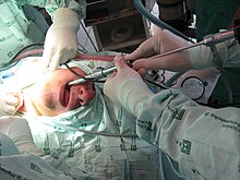
Rigid bronchoscopy
Rigid bronchoscopy is performed under anesthesia. The risk of injury is higher than with flexible bronchoscopy, but the larger lumen also allows more extensive interventions. It is the preferred procedure for persistent pulmonary bleeding and bronchoscopy in children. In addition, it is often used in the diagnosis of lung tumors , the removal of foreign bodies, the use of laser scalpels or argon-ion lasers and the insertion of a stent into the trachea.

Flexible bronchoscopy
Flexible bronchoscopy has largely replaced rigid bronchoscopy in the daily routine. With the exception of a few special applications, all rigid bronchoscopy tasks can be achieved faster, more conveniently and more gently for the patient (no anesthesia) with the flexible instruments. Flexible bronchoscopes now only have a diameter of two to three millimeters and can therefore also be used with very young children. They can be inserted deeper than a rigid bronchoscope, have a lower risk of injury and the patient can be awake or only slightly sedated.
- Diagnostic use at
- Look for lung tumors
- Taking sample material, biopsy , performing a bronchial lavage , taking samples for microbiological testing for germs (bacteria, fungi, parasites).
- Planning the local treatment of a tumor, local radiation therapy
- Diagnostics of foreign bodies, whereby their removal still requires the additional use of rigid instruments.
- Clarification of constrictions in the airways
- Determination of insufficient ventilation of the lungs ( atelectasis ), e.g. B. after a major operation
- Therapeutic use
- Irrigation and suction of a mucus plug from the bronchi, also with the ventilation hose ( tube ) lying down , see also: bronchoalveolar lavage
- Correction of the position of the ventilation hose
- Fiberoptic intubation or assistance with puncture tracheotomy

Bronchoalveolar Lavage (BAL)
Here, the bronchoscope is up in a segmental advanced so far and performed a rinse that the alveoli to be washed in a segment. This lavage liquid is examined for specific cells and ingredients.
Cryobiopsy
This technique is used within a bronchoscopy by cooling the tip of the deeply inserted instrument so much that lung tissue freezes to it and can be pulled out with the instrument. These tissue samples are often more informative in histological assessment than biopsies obtained with biopsy forceps.
literature
- Claus Kroegel, Ulrich Costabel: Clinical Pneumology: The reference work for clinics and practices . Georg Thieme, Stuttgart 2014, ISBN 978-3-13-129751-8 , pp. 133-134 .
Web links
Individual evidence
- ↑ Venerino Poletti, Gian Luca Casoni, Carlo Gurioli, Jay H. Ryu, Sara Tomassetti: Lung cryobiopsies: A paradigm shift in diagnostic bronchoscopy ?. In: Respirology. 19, 2014, p. 645, doi : 10.1111 / or 12309 .
