Atelectasis
| Classification according to ICD-10 | |
|---|---|
| P28.0 | Primary atelectasis in the newborn |
| P28.1 | Other and unspecified atelectasis in the newborn |
| J98.1 | Lung collapse (including atelectasis) |
| ICD-10 online (WHO version 2019) | |
Under an atelectasis (from Greek ἀτελῆς, ateles "incomplete" ( alpha privativum and τελος, telos "end," goal "); ἐκτάσις (from εξ, ex- or ek-" from, from ", and τασις, tasis" stretching " ), ektasis " expansion ") is understood as a collapsed section of the lung that is filled with little or no air. The alveolar walls lie against one another.
to form
Congenital atelectasis
Congenital atelectasis is also known as primary atelectasis or fetal atelectasis . The lungs are not or only incompletely ventilated after birth . The possible causes are central respiratory disorders (e.g. damage to the respiratory center ), amniotic fluid aspiration , lung malformations and lung immaturity ( surfactant deficiency).
A special form of primary atelectasis is a typical finding in a stillbirth : Since the lungs are physiologically unfolded in fetal life , but contain no air (but amniotic fluid), this finding can also be used for forensic medicine in order to prove that breathing activity is present after birth or exclude.
Acquired atelectasis
Acquired atelectasis is also known as secondary atelectasis . All acquired atelectases have minimal residual air in the atelectatic lung segments and are in principle reversible.
- Relaxation atelectasis: After injury to the chest wall, air enters the pleural space ( pneumothorax ). The lung is no longer held against the chest wall and collapses.
- Compression atelectasis: Due to pleural effusion , tumors of the pleura or mediastinum as well as enlarged mediastinal lymph nodes , due to thoracic deformities . Sections are poor in air and blood.
- Resorption atelectasis: In the case of ventilation with 100% oxygen, the N 2 otherwise present in the alveoli is missing . When all of the O 2 diffuses into the capillaries, the alveoli collapse. Physiologically, they are kept open by nitrogen.
- Obstructive atelectasis: after a bronchus has been relocated , e.g. B. by purulent bronchitis in chronic bronchitis , enlarged hilar lymph nodes (e.g. in tuberculosis , sarcoid or metastases ), tumors or foreign bodies, the distal air is trapped and absorbed. The portion of the lung located in the supply area of the bronchus shrinks.
- Microelectasis: Atelectasis caused by insufficient breathing or insufficient ventilation volume , which is only shown by an elevated diaphragm in the X-ray image.
In intensive care patients, atelectasis is the most common pulmonary complication after surgery.
Diagnosis

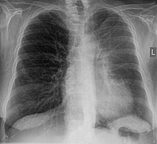
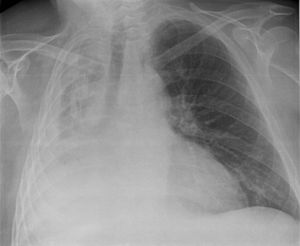
With the percussion you can hear a dampening of the lung sound. On palpation there is a weakening of the voice fremitus . The sound of breathing is weakened during auscultation of the lungs . In the chest x-ray, the direct signs are a decrease in transparency and displacement of the clefts of the lobes, and indirect signs include an elevated diaphragm , mediastinal displacement , compensatory emphysema , hilar displacement and narrowing of the ribs. Pneumonia must be considered in the differential diagnosis ; in contrast to this, atelectasis does not show a bronchopneumogram . As an alternative or in addition to the X-ray, a computed tomography image can be made. In ultrasound to atelectasis seen in the area of pleural effusion as a non-ventilated, volume decreased lung sections.
consequences
Most atelectases resolve on their own or are eliminated by medical and nursing measures. The complication can be inflammation and edema , later fibrosis . Depending on the size of the atelectasis, the surface area for gas exchange is reduced, which can lead to a right-left shunt , hypoxia and central cyanosis . In the atelectatic segment of the lung, blood flow to the lungs is reduced by vasoconstriction ( Euler-Liljestrand mechanism ). This increases the resistance in the pulmonary circulation and thus the load on the right heart . It can cause a pulmonary heart come. Pneumonia (pneumonia) can develop in the non-ventilated parts of the lungs .
therapy
Physical measures (change of position, cold water applications, tapping massages, breathing exercises) are very effective. The effect of mucolysis (= liquefaction of mucus) is controversial. Pain management is necessary for rib fractures or pleurisy . Theophylline , beta sympathomimetics and antibiotics can be used as medication for bacterial infection. The pus or viscous secretions and foreign bodies are removed by means of bronchoscopy . The use of PEEP or CPAP has also proven itself as an additional therapy strategy for reopening (recruiting) and keeping affected alveoli open .
In atelactesis-related hypoxic lung failure, mechanical, if possible non-invasive , ventilation is used.
literature
- Müller, Fraser, Colman, Paré: Radiologic Diagnosis of Diseases of the Chest. WB Saunders Company, 2001, ISBN 0-7216-8808-X .
- Wegener: whole body computed tomography. Blackwell Wissenschaft, Berlin 1992, ISBN 3-89412-105-X .
- Wolfgang Oczenski: breathing - breathing aid: breathing physiology and ventilation technology. 9th edition. Georg Thieme Verlag, 2012, ISBN 978-3-13-137699-2 .
- Rudolf Frey : Lung atelectasis and collapse. In: Ludwig Heilmeyer (ed.): Textbook of internal medicine. Springer-Verlag, Berlin / Göttingen / Heidelberg 1955; 2nd edition, ibid. 1961, pp. 709-712.
Web links
Individual evidence
- ↑ Hans Jantsch, G. Lechner: Radiological monitoring of the ventilated patient. In: J. Kilian, H. Benzer, FW Ahnefeld (ed.): Basic principles of ventilation. Springer, Berlin a. a. 1991, ISBN 3-540-53078-9 , 2nd, unchanged edition. Ibid 1994, ISBN 3-540-57904-4 , pp. 134-168, here: p. 162.
- ↑ Hans Jantsch, G. Lechner: Radiological monitoring of the ventilated patient. In: J. Kilian et al. (Ed.): Basic principles of ventilation. 2nd, unchanged edition. Berlin et al. 1994, pp. 134-168, here: pp. 159-163.
- ^ Klaus Holldack, Klaus Gahl: Auscultation and percussion. Inspection and palpation. Thieme, Stuttgart 1955; 10th, revised edition, ibid 1986, ISBN 3-13-352410-0 , p. 90 f.
- ^ Rolf Dembinski: Non-invasive forms of ventilation. In: Anesthesia & Intensive Care Medicine. Volume 60, June 2019, pp. 308-315, here: p. 309.
- ↑ Christian Putensen, Thomas Muders, Stefan Kreyer, Hermann Wrigge: Alveolar ventilation and recruitment under lung protective ventilation. In: Anaesthesiology, Intensive Care Medicine, Emergency Medicine, Pain Therapy. Volume 43, No. 11/12, 2008, pp. 770-777.
- ^ Rolf Dembinski: Non-invasive forms of ventilation. In: Anesthesia & Intensive Care Medicine. Volume 60, June 2019, pp. 308-315, here: pp. 309, 312 and 314.


