Shunt (medicine)
A shunt (English for “shift”, “shunt”, “soft”; pronunciation: [ ʃʌnt ]) is a short-circuit connection with fluid transfer between normally separated vessels or cavities. This can occur naturally (e.g. in the context of malformations such as arteriovenous malformations ) or it can be artificially applied as part of a medical measure.
Information on therapy can be found under the keywords atrial septal defect and ventricular septal defect .
Embryonic Shunts
There are three shunts during the child's embryonic and fetal period. The liver shunt, the aortic shunt and the atrial shunt. These three shunts are necessary in the embryonic circulation because the child cannot supply himself with oxygen by breathing. As long as the lungs are collapsed, the child relies on the oxygen provided by the placenta. The shunts close after birth. However, failure to seal can cause various heart defects that can be surgically corrected.
Liver shunt
The liver shunt is characterized by the fact that the umbilical vein (vena umbilicalis) bypasses the liver circulation, in which it connects through the ductus venosus (also called ductus arantii) with the inferior vena cava (inferior vena cava) and thus goes directly to the heart. As a result, mixed blood, i.e. low-oxygen (from the inferior vena cava) and high-oxygen (from the umbilical vein), arrives in the child's heart. The oxygen and nutrient content of the blood of the umbilical vein is, however, reduced far less than it would be without the shunt due to the liver shunt, so that the oxygen content of the mixed blood is still sufficient for the child's supply.
Atrial shunt
During the development of the heart, the heart tube is divided cranially by the heart septa along the blood flow path. This division by tissue is called septation. First a septum primum arises, which divides the common atrium into a right and a left. With this atrial division, however, a foramen primum (“first hole”) remains . This hole is near the endocardial cushion, which lies medially in the heart. This hole closes and a new hole is created: the foramen secundum or foramen ovale . It is important that the foramen ovale is formed in the septum primum. The foramen ovale is closed by a kind of curtain, the septum secundum . This septum arises on the right half of the heart, so it belongs to the right atrium. This septum guarantees the flow of blood from the right atrium to the left, preventing blood exchange from the left atrium to the right. The foramen ovale thus forms the atrial shunt to guide the mixed blood from the inferior vena cava into the left atrium so that the left heart can supply the body with more oxygenated blood.
Aortic shunt
Since the lungs are not yet functional and have collapsed, the ductus arteriosus (or Ductus Botalli) bypasses the lungs . Starting from the pulmonary trunk, the ductus botalli goes to the aorta . However, the ductus botalli only leads the deoxygenated blood from the right ventricle to the aorta when the common carotid artery and the subclavian artery have departed. Thus, the brain and the arms get the most oxygenated blood, the rest of the body is supplied with less oxygenated blood.
Hemodynamic Shunts
Symptoms
- A hemodynamic shunt (or circulatory shunt ) can significantly change the pressure conditions in the blood vessels . For example, a shunt in the context of a ventricular septal defect can limit the achievable blood pressure.
- A shunt can also reduce the oxygen saturation of the blood through mixing. Since the relationship between saturation and partial pressure is not linear , but sigmoid , this can result in an undersupply of the organism.
Shunt between large and small circulation
In the case of circulatory shunts, a distinction is made between a left-right shunt , in which arterialized blood flows back into the venous system, bypassing the peripheral capillaries, and a right-left shunt , in which oxygen-poor blood enters the body arteries.
Such short-circuit connections between the pulmonary circulation emanating from the right half of the heart and the body circulation emanating from the left half of the heart are clinically significant in the case of congenital heart defects ( atrial septal defect and ventricular septal defect ) or in other developmental disorders such as e.g. B. the persistent ductus arteriosus (open ductus arteriosus Botalli) or a persistent foramen ovale (PFO). According to the higher pressure in the body circulation of the left heart, there is always a left-right shunt; If the permanent stress on the right heart results in a structural change in the same, a shunt reversal with a subsequent right-left shunt can occur. This process is called the Eisenmenger reaction (with resulting Eisenmenger syndrome ).
With the right-left shunt, the cardiac output is greater than the lung output ; with the left-right shunt, the cardiac output is smaller than the lung output. The shunt volume is the volume of blood flowing through a shunt per unit of time. In general, the following applies to bidirectional shunts : cardiac output + left-right shunt volume = lung time volume + right-left shunt volume. This is why the following applies to the left-right shunt, if the right-left shunt volume is zero: "Large minute volume in the small circuit → small minute volume in the large circuit".
In the physiology of respiration, a distinction is made between the following types of shunt. Examples:
- Physiological shunt: the venous admixture in oxygenated blood
- Anatomical or pathological shunt: venous admixture from blood vessels between the large and small blood circulation
- Functional shunt: venous admixture from unventilated or poorly ventilated alveoli
- intrapulmonary shunt between veins and arteries in the pulmonary circulation bypassing the alveoli
Congenital arteriovenous shunts of the peripheral vessels are called AV malformation . This is a congenital vascular connection between an artery and a vein without a capillary bed in between . Iatrogenically , short-circuit connections ( AV fistula ) can also accidentally arise when an artery is punctured through a vein, for example during a cardiac catheter examination .
Shunt surgery on the heart
In the case of certain congenital heart defects, an artificial shunt is placed between the arterial and venous circulation to improve the oxygen supply to the patient. The classic shunt is the Blalock-Taussig anastomosis . In many cases this shunt is removed again as part of a corrective or further palliative operation for the heart defect.
Dialysis shunts
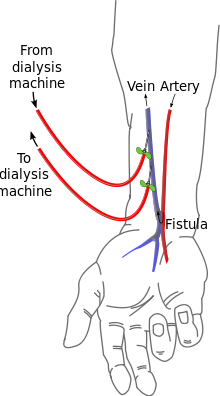
In dialysis patients , a shunt (or arteriovenous fistula ) is artificially created in order to have a large-volume vessel available with which hemodialysis can be performed. This method was developed by Belding Scribner in 1960 .
The preferred location for a dialysis shunt is the connection between the radial artery and the cephalic vein on the forearm. This shunt is also called the Cimino-Brescia shunt after its first description . Other options for a dialysis shunt on the upper arm are the connections between the mobilized basilica vein and the brachial artery (known as the basilica shunt ) or between the cephalic vein and the brachial artery (known as the cephalic shunt ).
Dialysis shunts are vital for patients with severe renal impairment ( renal insufficiency ).
When shunts are infected, specific or empirical antimicrobial therapy is used.
Ventriculo-peritoneal shunt
In neurosurgery, a so-called VP shunt is a permanent drainage of the liquor in the case of hydrocephalus , which is surgically applied. A tube is led from the top of the skull under the skin through the neck down in front of the chest wall to the peritoneum cavity ( peritoneal cavity ). There are VP shunts that have a valve that can be adjusted from the outside in order to adapt the discharge to requirements (see also intracranial pressure ). A further article on cerebral shunts is devoted to this topic in detail.
literature
- Lutz Steinmüller, Marc Olaf Liedke, Margret Liehn: Shunt and port systems. In: Margret Liehn, Brigitte Lengersdorf, Lutz Steinmüller, Rüdiger Döhler (eds.): OP manual. Basics, instruments, surgical procedure. 6th, updated and expanded edition. Springer, Berlin / Heidelberg / New York 2016, ISBN 978-3-662-49280-2 , pp. 321–327.
Web links
Individual evidence
- ^ Stefan Silbernagl , Florian Lang: Pocket Atlas of Pathophysiology. Georg Thieme Verlag, Stuttgart / New York, ISBN 3-13-102191-8 , and Deutscher Taschenbuch-Verlag, Munich 1988, ISBN 3-423-03236-7 , p. 202.
- ↑ Willibald Pschyrembel : Clinical Dictionary . 266th edition. Verlag Walter de Gruyter, Berlin 2014, ISBN 978-3-11-033997-0 , p. 1958
- ↑ Otto Martin Hess, Rüdiger WR Simon: Heart catheter: Use in diagnostics and therapy . Springer, 2013, ISBN 978-3-642-56967-8 , pp. 17 ( google.de ).
- ^ Günter Thiele (ed.): Handlexikon der Medizin . Urban & Schwarzenberg, Munich / Vienna / Baltimore 1980, 4 volumes, volume 4, p. 2241
- ↑ Gerd Herold : Internal Medicine 2019. Cologne 2018, p. 186.
- ↑ Thomas Pasch, S. Krayer, HR Brunner: Definition and parameters of acute respiratory insufficiency: ventilation, gas exchange, respiratory mechanics. In: J. Kilian, H. Benzer, FW Ahnefeld (ed.): Basic principles of ventilation. Springer, Berlin a. a. 1991, ISBN 3-540-53078-9 , 2nd, unchanged edition, ibid 1994, ISBN 3-540-57904-4 , pp. 95-108; here: pp. 98-100.
- ^ Marianne Abele-Horn: Antimicrobial Therapy. Decision support for the treatment and prophylaxis of infectious diseases. With the collaboration of Werner Heinz, Hartwig Klinker, Johann Schurz and August Stich, 2nd, revised and expanded edition. Peter Wiehl, Marburg 2009, ISBN 978-3-927219-14-4 , p. 72 f. ( Shunt infections ).
